Orofacial Pain Referral: https://thinkbetterlife.com/referrals/
View patient video testimonials: https://www.youtube.com/channel/UCk9Bfz6pklC7_UluWFHzLrg/videos
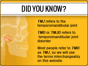
What is Orofacial Pain and how does it relate to TMJ Disorders and Headaches.
 Please visit this entire site to learn about help available for treating your Orofacial Pain and visit the patient testimonials about how they received help with their Orofacial Pain.
The term Craniofacial pain, is also commonly used by dental groups as well as universities such as Tufts.
THERE ARE MANY DIFFERENT WAYS TO APPROACH IMPROVING YOUR QUALITY OF LIFE AS RAPIDLY AS POSSIBLE. RELIEF OF PAIN IS ALWAYS THE FIRST STEP OF REHABILITATION. NEUROPLASTICITY IS THE PROCESS WHERE CONTINUED PAIN CAN BECOME CHRONIC PAIN THROUGH THE PROCESS OF NEUROPLASTICITY. THIS IS THE BIGGEST DANGER WHEN DEALING WITH OROFACIAL PAIN!
According to the Academy of Orofacial Pain the term covers the following subjects that I have expanded on:
1. TMJoint Disorders or TemporoMandibular disorders. These can be internal derangements or intracapsular disorders, capsular problems and extracapsular problems.
2. Pain in the Masticatory Muscles including but not limited to Myofascial Pain and Dysfunction
3. Cervical musculoskeletal pain also including but not limited to Myofascial Pain and Dysfunction
4. Neurovascular pain: I have gone into great lengths discussing the Trigeminovascular system and the trigeminal cervical complex in previous posts describing the role of the trigeminal nerve in all headaches.
5. Neuropathic pain
6. Sleep disorders related to orofacial pain which would include sleep apnea, snoring bruxism, nocturnal clenching and other movement disorders or parasomnias
7. Orofacial Dystonias The Dystonia Society website describes these a:
“In oromandibular dystonia the muscles that move the mouth and jaw are affected by involuntary spasm. This unwanted muscle contraction can pull the mouth and/or tongue into different positions. This often happens when people are using their mouths e.g. talking or eating, but can happen at rest as well.Like most types of dystonia it can be made worse when people are anxious or tired. It does not affect the mind or senses.
Although oromandibular dystonia most commonly develops following spread of dystonia from the neck or eyes, it can also appear in isolation. Where the condition comes on in mid-life without obvious cause, it will not usually spread further.
In some people, previous treatment with medicines that work by blocking the chemical dopamine in the brain (which can be used to treat a variety of conditions including nausea, vertigo or anxiety as well as psychiatric conditions such as schizophrenia and depression) can be the cause of oromandibular dystonia.”
Dr Brendan Stack has done very interesting work with changing maxillo -mandibular relations in many patients with movement disorders utilizing non-drug treatment regimens. I strongly Rx patients with these disorders at least become aware of the work Dr Stack is doing.
8. Headaches, Migraines ,autonomic cephalgias and various types of referred pain to the head. There is obvious overlap to neurovascular pain and referred cervicall pain.
Intraoral, intracranial, extracranial, and systemic
According to the AOP website “The field of Orofacial Pain is concerned with the prevention, evaluation, diagnosis, treatment, and rehabilitation of orofacial pain disorders. Such disorders may have pain and associated symptoms arising from a discrete cause, such as postoperative pain or pain associated with a malignancy, or may be syndromes in which pain constitutes the primary problem, such as TMJ disorder pain, neuropathic pains or headaches.”
THE FOLLOWING SECTION WILL COMMENT ON AN ARTICLE IN THE JOURNAL OF PAIN RESEARCH FROM THE US LIBRARY OF MEDICINE , NATIONAL INSTITUTE OF HEALTH. THERE IS NOT UNIVERSAL AGREEMENT ABOUT THE BEST TREATMENT FOR OROFACIAL PAIN. I OFFER ADDITIONAL PERSPECTIVES AND INFORMATION ON TREATMENT GOALS AND QUALITY OF LIFE CONSIDERATIONS.
***ALL MY COMMENTS WILL BE IN ALL CAPITALS!******MANY WILL BE IN BOLD
Orofacial pain management: current perspectives
Disorders of the TMJ ****I PREFER THE TMJoint WHICH CLEARLY EXPLAINS WE ARE DISCUSSING ACTUAL JOINT ISSUES
Disorders of the TMJ **TMJoint** are a result of a disc–condyle incoordination that influences the TMJ biomechanics. These disorders comprise the disc interference disorders or internal derangements, such as disc displacements with and without reduction, that can be asymptomatic or symptomatic due to inflammation THEY ARE ALWAYS SYMPTOMATIC BUT NOT ALWAYS PAINFUL BUT BECOME PAINFUL WHEN THE LEVEL OF INFLAMATION INCREASES. INFLAMATION CREATES AN ENVIRONMENT FOR ADDITIONAL JOINT INJURY. (eg, capsulitis/synovitis). Disc displacements with reduction may present as a painful or non-painful click. REDUCTION REFERS TO REDUCING OF THE DISLOCATION OR RETURN TO HEALTHIER CONDITION. Disc displacements without reduction may present with a painful limitation at opening. IDEALLY, PATIENTS CAN BE PREVENTED FROM DEVELOPING DISPLACEMENTS THAT ARE INCREASING IN DAMAGE, PAIN AND INFLAMATION. Retrodiscitis and TMJ subluxation (CONDYLE DISLOCATION OVER EMINENCE) may present symptomatology when the pain is a result of inflammation arising from the retrodiscal tissues or capsulitis or synovitis processes. RETRODISCAL TISSUES BECOME INFLAMED AND PAINFUL DUE TO IMPROPER PHYSIOLOGICL FUCTION WITHIN THE TMJoint. Osteoarthritic changes can originate in the TMJ articular surfaces and, when they are influenced by a systemic disease, can become aggressive and progressive, such as in the case of polyarthritis. THIS IS TRUE OF ALL JOINTS IN THE BODY BUT THE TMJoint IS UNIQUE IN THAT IT CAN OFTEN BE STABILIZED TO PREVENT FUNCTIONAL CHANGES AND RESULTANT INFLAMMATORY ARTHRITIS.
Muscular disorders
Myalgia THE TERM MYALGIA SPECIFICALLY REFERS TO MUSLCE PAIN NOT WHY THERE IS MUSCLE PAIN usually presents as a dull aching pain due to muscle injury MICRO INJURY AND REPETITIVE STRAIN ARE VERY COMMON or strain. It is commonly seen in acute forms THE MOST COMMON PRESENTATION IS MYOFASCIAL PAIN AND DYSFUNCTION AS DESCRIBED BY DR JANET TRAVELL, though, with continued muscle tension,CHRONIC MUSCLE SHORTENING LEADS TO PHYSIOLOGIC CHANGES IN THE MUSCLE FIBERS INCLUDING TAUT BANDS AND TRIGGER POINTS can present for longer periods of time. Treatment may include, rest, hot or cold compresses, stretching exercises, and muscle relaxants. MYOFASCIAL PAIN IS MYALGIA OR MUSCLE PAIN Myofascial pain (MFP) also presents as a dull, continuous aching pain that varies in intensity. MFP produces pain upon palpation that is local and may refer to other sites, as mapped out by TRAVELL & Simons et al. MFP tends to be seen in muscle pain conditions of a more chronic nature, DOES THIS MEAN THAT LACK OF TREATMENT EARLY RESULTS IN MYOFASCIAL PAIN FORMATION? in which the tension is unremitting. TRIGGER POINTS IN MYOFASCIAL PAIN ARE NOT NON-REMMITING BUT ACTUALLY GO THROUGH TWO PHASES LATENT AND ACTIVE WHERE ACTIVE CREATES MUCH MORE PAIN AND PASSIVE IS PAINFUL ON PALATION BUT IS STILL A PHYSIOLOGICAL PROBLEM. Trigger points can often be USUALLY seen in MFP and may be localized to a taut band of muscle. In addition, trigger points are associated with decreased muscle length and, when stimulated, can result in a local twitch response. THIS IS DUE TO AN AUTONOMIC DYSFUCTION WITHIN THE MUSCLE RELATING TO THE MUSCLE SPINDLES Palpation of the trigger points should duplicate the patient’s pain complaint, THIS IS ONLY TRUE WHEN THE TRIGGER POINT IS IN AN ACTIVE PHASE BUT THEY DO NOT CAUSE REFERRED PAIN IN LATENT PHASE. thus confirming diagnosis. Blocking the source of the pain (ie, masseter muscle) by using a vapocoolant spray or local anesthetic injection can also provide a definitive diagnosis. THE USE OF VAPOCOOLANT SPRAY AS DESCRIBED BY TRAVELL IS ACTUALLY A TREATMENT OF THE TAUT BAND AND TRIGGER POINT. DEACTIVVATION AND ELIMINATION OF TRIGGER POINT IS ACCOMPLISHED BY UTILIZING TECHNIQUES DESCRIBED IN THE PAIN GATE THEORY, THE COLD SPRAY FLOODS THE AFFERENT NERVE INPUT BLOCKING THE PAIN OF THE STRETCH ALLOWING BREAKING UP OF TAUT BANDS BY BLOCKING ACTION OF MUSCLE SPINDLES TO PROTECT AREA OF PAIN.
Myositis is a localized transient swelling involving the muscle and facial tissues. THERE ARE BOTH SYSTEMIC AND LOCAL CAUSS OF MYOSITIS WHICH IS TYPICALLY AN INFLAMATORY RESPONSE WITHIN THE MUSCLE TISSUE CREATING PAIN AND DECREASED FUNCTION. There tends to be increased pain with mandibular movement and localized tenderness, usually following injury or infection. LOCAL MYOSITIS OFTEN OCCURS FOLLOWING INFERIOR ALVEOLAR NERVE BLOCKS AND LOWER THIRD MOLAR REMVAL AS WELL AS BEING RELATED TO EXTENDED DENTAL APPOINTMENTS REQUIRING WIDE MOUTH OPENING.
Patient evaluation
TMD assessment should include a general examination of the head and neck, a detailed examination of the masticatory muscles,AS WELL AS CERVICAL AND POSTURAL MUSCULATURE, EVALUATION OF SUPRA NA INFRAHYOID MUSCLES AND SPECIAL ATTENTION TO THE SCALENE GROUP OF MUSCLES DUE TO THEIR IMPORTANCE IN BOTH POSTURE AND BREATHING an evaluation of the TMJs, an evaluation of mandibular range of motion (ROM), and a detailed intraoral examination.13
Evaluation of the TMJs and mandibular ROM
The evaluation of the TMJs includes examination for any signs of dysfunction or pain symptomatology. Fingertips are placed over the lateral and posterior aspects of the TMJs applying light but steady force and performed when the mandible is at rest/closed position and opening. Symptomatology reported in response to force applied to the lateral aspect of TMJs may be a sign of capsulitis/synovitis. Symptomatology reported in response of force applied to the posterior aspect of the condyle may be a sign of retrodiscitis or posterior capsulitis. AN EXCELLENT METHOD OF EVALUATING THE TMJoints IS THE INTRA-MEATAL EXAMINATION WHERE POSTERIORIZATION OF THE CONDYLE INTO THE EAR CANAL CAN BE EASILY PALPATED. I FREQUENTLY HAVE PATIENTS DO THIS WHITH THEIR PINKIES SO THEY CAN FEEL THE TISSUE DISPLACEMENT INTO THE EARS. DO THE CONDYLES BELONG IN THE EARS? COSTEN, THE ENT WHO ORIGINALLY DESCRIBED TMJoint DISORDERS WAS WELL AWARE OF THE POSTERIOR DISPLACEMENT ESPECIALLY IN EDENTULOUS AND DENTURE PATIENTS. WHEN COSTEM DID HIS EXAMS PARTIAL AND COMPLETE EDENTULISM OCCURED AT A MUCH YOUNGER AGE THEN TODAY.
The clinician should be aware of joint sounds, which could present as clicks, pops, or crepitus. CREPITUS IS THE SOUND SIMILAR TO SAND OR BROKEN GLASS IN A JOINT FROM BONE ON BONE CONTACT These sounds are evaluated with the help of a stethoscope placed in the TMJ area or sometimes perceived during palpation. SONOGRAPHY AND JOINT VIBRATIONAL ANALYSIS CNA QUANTIFY THESE SOUNDS. SPECTRAL ANALYSIS OF THESE SOUNDS CORRESPONDS WITH FINDINGS ON MRI AND CAT SCANS. Clicks and pops are commonly related to disc displacements with reduction and crepitation is commonly associated with osteoarthritic changes in the articular surfaces of the TMJ. THIS SIMPLE STATEMENT IS THE RATIONALE FOR EARLY NON-INVASIVE TREATMENT TO PREVENT FURTHER DEGRADATION OF THE TMJoint. Imaging of the TMJ may also be useful during examination. Moreover, it is very important to identify any TMJ restrictions. The clinician should view the patient’s opening and closing patterns to note any mandibular deviations. THIS CAN BE MEASURED MORE ACCURATELY AND QUANTIFIED WITH VARIOUS COMPUTERIZED SCANS SUCH AS THOSE AVAILABLE BY MYOTRONICS AND BIORESEARCH. The evaluation of mandibular ROM consists of measuring comfort opening, active opening, passive opening, protrusion, and left and right lateral excursions with a millimeter ruler COMPUTERIZED TESTING IS MORE REVEALING AND ACCURATE while noting the severity and location of pain with jaw movement. This can be particularly helpful in differentiating between joint and muscle pain. FREQUENTLY THERE IS BOTH MUSCLE AND JOINT PAIN OCCURING SIMULTANEOUSLY Comfort opening is determined by the patient opening as wide as possible without any pain, active opening is determined by the patient opening as wide as possible with pain, and passive opening is determined by the clinician gently stretching the patient presumably past active opening while noting a soft or hard end feel. PAIN ASSOCIATED WITH JOINT NOISES SHOULD BE CONSIDERED VERY IMPORTANT A reasonably normal interincisal distance is approximately 40 mm,THIS VARIES BY SEX OF PATIENT AND OVERALL SIZE OF PATIENT or the width of three of the patient’s fingers as a crude measure. Usually, with proper questioning, the patient will reliably reveal any recent limitations in ROM. The occurrence of TMJ clicking, crepitus, or jaw opening interferences with or without pain should also be noted at the initial examination. These baseline findings ARE FAR MORE ACCURATE WHEN TAKEN WITH BIOMEDICAL INSTRUMENTATION will aid in establishing the differential diagnosis and treatment options, as well as providing a comparison for future change in TMD symptoms.
Evaluation of the muscles of mastication
The muscles of mastication should be palpated bilaterally for firmness and tenderness, utilizing approximately 2–3 lbs of pressure, or the amount of pressure needed to cause blanching of the fingernail. Upon muscular palpation, the patient should be asked to report the severity of the tenderness, pain referral to multiple sites or single-site pain localization, and replication of the chief complaint upon palpation. It may be pertinent to ask the patient about their use of analgesics prior to palpation in order to account for reduced symptoms upon examination. IT IS ESSENTIAL TO MAP OUT REFERRAL PATTERNS AND TO PALPATE BOTH ORGIN, INSERTIONS AND BODIES OF EACH MUSCLE. THE POSTURE OF THE PATIENT WILL AFFECT THE RESPONSE TO PALPATION. THIS PART OF THE EXAM IS INCREDIBLE IMPORTANT IN UNDERSTANDING THE UNDERLYING CAUSES OF PAIN. SURFACE AND NEEDLE EMG BBOTH HAVE A PLACE IN DIAGNOSIS AND TREATMENT IN SOME PATIENTS. SURFACE EMG IS ESPECIALLY REVEALING NON-SYMETRICAL RESTING VALUES AND FUNCTIONING MUSCLE ACTIVITY.
Management of TMD
Most of the time, patients will visit the clinician when pain and dysfunction, such as limitation of opening, episodes of joint locking (open lock/TMJ subluxation), pain with mandibular function (chewing), facial pain, or headache are present.
The treatment goals for TMD are decreasing pain, restoring normal ROM, and restoring normal masticatory and jaw function. Many TMDs can be cyclical and self-limiting, with periods of complete remission of symptoms. A MAJOR GOAL OF THE CLINICIAN SHOULD BE TO CLEARLY EXPLAIN THE FUNCTIONAL ETIOLOGIES OF THESE PROBLEMS AND THE BEST AVAILABLE CARE NOT JUST TREAT THE SYMPTOMS
In the case of disc-condyle LACK OF coordinations, studies suggest that for some patients even though they may be progressive (for example, a disc displacement with reduction may progress to a disc displacement without reduction), they are self limiting, suggesting an adaptation of the condition and with no significant disability. DISABILITY IS IN THE EYES OF THE PATIENTS. THE IDEA THAT LIVING IN PAIN FOR AN EXTENDED TIME IS SELF LIMITING IS A LIE. LIVING IN PAIN CAN BECOME A CHRONIC PAIN PROBLEM AND HAVING MANY EMOTIONAL AND SOCIAL EFFECTS FAR BEYOND THE CLICKING OR JOINT PAIN. It is very important to emphasize that patients have no recurrence of symptoms with the use of conservative, reversible treatments, IS CONSERVATIVE TREATMENT ONE THAT ALLOWS CONTINUAL DESTRUCTION OF THE JOINTS EVEN IF PAIN IS NOT CONSTANT? thus conservative treatment is the modality that needs to be used at all times. CONSERVATIVE TREATMENT SHOULD BE USED AT ALL TIME IS A STATEMENT THAT CAN BE VERY DAMAGING TO THE HEALTH OF PATIENTS. Initial treatment should therefore stress a conservative ABSOLUTELY and reversible approach. Primary treatment options include home care (self-care program), medical care (non-surgical care), and surgical care. SURGERY IS ALMOST ALWAYS BEST AVOIDED. IF CONSERVATIVE NON-REVERSIBLE TREATMENT CAN PREVENT THE NEED FOR FUTURE INVASIVS AND POSSIBLY DISABLING SURGERY SHOULD THE PATIENT ALWAYS BE PRESENTED WITH LONG TERM TREATMENTS THAT MAY PREVENT FURTHER JOINT DEGRADATION.
Patient education: home care program
Home care I WOULD PREFER TO SAY EDUCATION OF THE PATIENT should generally be INCLUDED the initial approach, at least as part of a more extensive treatment plan. The use of a home care program has proved to be effective in the management of TMD. It has been shown that patients have reported feeling less pain immediately after their initial patient education/counseling visit, perhaps as a consequence of an immediate reduction in stress/tension-related parafunctional activity. THERE IS A NATURAL PROGRESSION OF THE DISEASE AND RANDOM FLUCTUATIONS OF INCREASED AND DECREASED PAIN FREQUENTLY OCCUR WITH OR WITHOUT INTERVENTION. Patient education is a crucial aspect of home care and is one of the most subtle and underappreciated, yet effective, treatments for TMD. Therefore, informing and reassuring the patient regarding their condition and presenting symptoms may alleviate a great deal of anxiety and improve treatment outcomes. THIS DOES NOT RELIEVE THE DOCTOR OF EXPLAINING POSSIBLE ADVERSE OUTCOMES FROM LACK OF MORE DEFINITIVE TREATMENTS.
A successful home care program consists of resting the masticatory muscles by limiting jaw movements, parafunctional habit modification, emphasizing a soft diet, and moist heat and/or ice therapy. IT IS INTERESTING THAT ELSEWHERE IN THE BODY ORTHOPEDIC PHYSICIANS AND PHYSICAL THERAPISTS FREQUENTLY UTILIZE LONG TERM ORTHOTICS AND PROSTHETICS TO TREAT MUSCLES AND JOINT BUT IN THE FAR MORE COMPLEX AND HIGHLY INNERVATED CRANIAL FACIAL REGIONS WE ARE MORE APT TO FOLLOW DEGENERATION RATHER THAN ATTEMPT TO PREVENT IT. Muscle rest may involve limited jaw activity (eg, reduced talking, chewing, yawning) for the treatment duration, and perhaps as a preventive measure, even after symptoms have resolved. DON’T USE YOUR JAWS IS NOT A CURE IT IS A DISABILITY, IDEALLY FULL FUNCTION SHOULD BE THE DESIRED OUTCOME. Patients with disc displacement without reduction should be instructed to avoid any forceful attempt to open the mouth wider when the condition is acute and have explained to them that, with the care provided, the ROM will improve and return to normal.
PHYSICAL THERAPY AND REHABILITATION IS BEST EMBARKED ON EARLY TO AVOID UTILIZING THE WRONG THERAPY OR THE RIGHT THERAPY AT THE WRONG TIME. PHYSICAL THERAPY SHOULD ALMOST ALWAYS PRECEDE SURGICAL INTERVENTION
Restricting the mandibular movements as much as possible would facilitate healing and prevent further injury. THIS IS NOT THE CASE IF THERE IS A CLOSE-LOCK DISLOCATION OR BLEEDING IN THE JOINT WHERE LIMITED MOVEMENT COULD LEAD TO ANKYLOSIS. This could be attained with a soft food diet, avoiding chewing gum and hard foods, and limitation of opening during yawning, as well as habit awareness, such as avoiding biting objects, clenching, or bruxing. Patients may have a diurnal (daytime) parafunctional habit (clenching, grinding, posturing) that is often not conscious, and this should be addressed to decrease sustained masticatory muscle contractions. MANY PATIENTS HAVE FUNCTIONAL PROBLEMS AND DEVIATE SWALLOWS THAT ACTUALLY PROTECT THE JOINT DURING SWALLOWING BUT LEAD TO OTHER PROBLEMS. WHEN ONE LOOKS AT THE ENTIRE SYSTEM IT FAILS AT THE WEAKEST POINT. THE WEAKEST POINT IS NOT ALWAYS EASILY ISOLATED. Patient education and understanding of the physiological rest position (lips together, teeth apart) is imperative in reducing and eventually halting the daytime activity that contributes to the progression of TMD. THE UTILIZATION OF AN ORAL MYOFUNCTINAL THERAPIST MAY BE THE MOST VALUABLE INTERVENTION FROM A BEHAVIORAL POINT OF VIEW. If asked to pay attention to their jaw position over time, many patients will return for follow-up with the recognition that they are in fact engaging in some jaw activity that contributes to their symptoms. OTHER PATIENTS MAY ACTUALLY FRE WORSE BECAUSE OF INCREASED OCCLUSAL AWARENESS THAT IS OFTEN A SIGNIFICANT FACTOR EFFECTING WELL BEING. Additionally, suggesting habit-controlling cues may be helpful in reminding the patient throughout the day to check the position of their bite. As an example, saying the letter “N” throughout the day can remind the patient to unclench or discontinue grinding their teeth. A soft diet is also crucial for muscle and TMJ pain management so that the condition is not exacerbated while treatment is provided. Finally, a trial of moist heat and/or ice therapy overlying the painful areas of the face, head, and neck can be recommended. Moist heat tends to work better for muscle pain or tension by increasing circulation and relaxing involved muscles, and ice for TMJ capsulitis by reducing inflammatory symptoms. UNFORTUNATELY NOT ALL PATIENTS RESPOND TO THESE TECHNIQUES AND MANY OFTEN CAN SUFFER ADDITIONAL INJURY FROM DELAYING APPLIANCE THERAPY.
Medical care (non-surgical)
Physical therapy
Instructing the patient to apply moist heat or cold compresses, or alternating both modalities, has been proven to be beneficial, since it stimulates analgesia and relaxation and may improve movement.
Physical therapy is beneficial in restoring the normal function of the TMJ, muscles of mastication, and cervical muscles, as well as in reducing inflammation, promoting repair, and strength. Physical therapy can be performed by an experienced physical therapist or can be provided by a qualified clinician who is treating the TMJ disorder. Primary goals of the physical therapy component of treatment are to stretch chronically contracted and fatigued muscles, increase ROM, and reduce muscular trigger point activity. A number of exercises are commonly used to treat TMJ-associated muscle disorders, including N-stretch (placing the tip of the tongue on the roof of the mouth and stretching the jaw) chin to chest (gently pulling the head forward, bringing the chin toward the chest); and head tilt (turning the head to one side and then tilting it posteriorly). These exercises must be done four to six times per day to be effective. In addition, the patient should use moist heat for 10–15 minutes followed by ethyl chloride spray prior to stretching the muscles. Vapocoolant spray provides a temporary anesthesia effect to the muscles so that a more intense stretch can be achieved without pain. THS IS ESSENTIAL IF THE TAUT BANDS AND TRIGGER POINTS ASSOCIATED WITH MYOFASCIAL PAIN ARE GOING TO BE SUCCESSFULLY TREATED. TRIGGER POINT INJECTIONS COMBINED WITH IMMEDIATE STRETCH PER TRAVELL AND PHYSICAL THERAPY IS EXTREMELY POWERFUL THERPY. YOUNGER PHYSICAL THERAPISTS ARE OFTEN TRAINED IN UTILIZING DRY NEEDLING AS PART OF PHYSICAL THERAPY PROTOCOLS . The heat and cooling spray should be used for at least three of the six exercising sessions throughout the day. THE BEST RESULTS ARE WHEN A HEATING PAD IS USED TO HEAT THE CORE (STOMACH) FOR 20 MINUTES PRIOR TO UTILIZING VAPOCOOLANT SPRAY. THE HEATING OF THE CORE CAUSES THE BODY TO SHUNT BLOOD TO THE FIVE LIMBS, HEAD, ARMS AND LEGS SO THE VAPOCOOLANT COOLS THE SKIN BUT NOT THE UNDERLYING MUSCLE. Patients can expect an even higher likelihood of treatment success if transcutaneous electrical nerve stimulation is added to a strict stretching regimen, and if biofeedback training is used as a cognitive behavioral procedure to teach the patient to maintain reduced muscular tension and pain. ULTRA LOW FREQUENCY TENS USED BY NEUROMUSCULAR DENTISTRY IS THE MOST EFFECTIVE AT ELIMINATING MUSCLE SPASM AND IS PRIMARILY A PERIFERAL EFFECT ON THE MUSCLES RATHER THAN PRIMARILY A CNS EFFECT WITH OTHER TENS.
Pharmacotherapy
Medications are an effective addition in managing the symptomatology of intracapsular disorders. Commonly used pharmacological agents for the treatment of TMJ disorders include analgesics, nonsteroidal anti-inflammatory drugs (NSAIDs), local anesthetics, oral and injectable corticosteroids, sodium hyaluronate injections, muscle relaxants, botulinum toxin injections, and antidepressants. The analgesics and corticosteroids are indicated for acute TMD pain; the NSAIDs, local anesthetics, and muscle relaxants are used for both acute and chronic conditions; and tricyclic antidepressants are usually used more for chronic TMD pain in association with tension-type headaches. THEY ARE ALSO VERY EFFECTIVE AT CONSOLIDATING DELTA SLEEP AND DELAYING REM SLEEP THAT IS HELPFUL IN TREATING MUSCLE PAIN FROM FIBROMYALGIA AND MYOFACIAL PAIN. THEY MAY BE HELPFUL IN DECREASING NOCTURNAL BRUXISM IN SOME PATIENTS. Research demonstrating the efficacy of botulinum toxin for muscular disorders related to TMD is limited, although there is some data to support the benefit of using low concentrations and large injection volumes of botulinum toxin at multiple muscular sites. THE MECHANISM OF BOTOX IS TO DECREASE THE NOCICEPTIVE INPUT TO THE TRIGEMINAL NERVOUS SYSTEM BY USING THE TOXIN TO PARALYZE THE NEUROMUSCULAR JUNCTION AND THEREFORE DECREASE PARAFUNCTION AND PAIN. CORRECTION OF NEUROMUSCULAR INPUT WITH A NEUROMUSCULAR ORTHOTIC IS SAFER AND MORE EFFECTIVE FOR MOST PATIENTS BUT BOTOX CAN BE USED ON AN INTERIM BASIS TO BREAK A PAIN CYCLE.
NSAIDs
NSAIDs are indicated for mild-to-moderate acute inflammatory conditions. Commonly used NSAIDs include ibuprofen and naproxen. NSAIDs should be used by the patient for a minimum of 2 weeks, with time-contingent usage as opposed to dosing based on the presence of pain. Long-term NSAID use is not recommended as long as the activity resulting in the inflammatory process can be reduced. In some chronic arthritic cases, the long-term use of NSAIDs, such as the COX-2 inhibitors, including celecoxib, may be considered; however, possible side effects, such as gastrointestinal upset, should be taken into account. IT IS ESTIMATED THAT AS MANY AS 40,000 DEATHS OCCUR ANNUALLY FROM SIDE EFFECTS OF NSAIDS.
Local anesthetics
Local anesthetics are primarily used when a myofascial trigger point is present. Myofascial trigger points are usually detected in the mastication muscles, but can also be found in numerous other muscles, such as the splenius capitis and upper trapezius. Due to its low toxicity to muscles, 1% procaine (1 cc) is recommended, but 1% or 2% lidocaine is also commonly used. THIS AUTHOR PRFERS THE USE OF 2% LIDOCAINE WITH NO PRESERVATIVES OR VASOCONSTICTORS. The trigger point injection technique involves locating the trigger point, which is usually found in a taut band of muscle, and needling the area. THE USE OF DRY NEEDLING VS LIDOCAINE INJECTION CAN VARY IN EFFECTIVENESS BY THE PATIENT. THE USE OF VAPOCOOLANT SPRAY CAN ELIMINATE THE PAIN FROM PENETRATION THROUGH THE SKIN. DR JANET TRAVELL IN HER LANDMARK TEXT MYOFASCIAL PAIN AND DYSFUNCTION : A TRIGGER POINT MANUAL CLEARLY STATES THAT INJECTION AND STRETCH IS VERY IMPORTANT, NOT JUST INJECTION. THE COMBINING OF ALL THREE MODALITIES WILL GIVE THE BEST RESULTS. The patient should be instructed that the muscles may be sore for the first 48 hours after the injection, but should begin to improve thereafter. I FIND THAT MOST PATIENTS HAVE IMMEDIATE RELIEF THAT IMPROVES OVER 24-72 HOURS AND SORENESS IS USUALLY FELT ONLY IF PRESSURE IS APPLIED OVER INJECTION SITES. The efficacy of trigger point injections is highly variable and dependent, for the most part, on the patient’s compliance with a strict physical therapy regimen in conjunction with the injections. MANY PATIENT ARE FAR MORE COMPLIANT WITH EXERCISE WHEN TRIGGER POINT THERAPY ELIMINATES MOST OF THE FUNCTIONL PAIN. In addition, local anesthetics can be used to block the likely source of pain to confirm a diagnosis.
TMJ injections
AN AURICULARTEMPORAL NERVE BLOCK CAN SIGNIFICANTLY REDUCE TMJoint PAIN AND CN BE USED DIAGNOSTICALLY AND AS A TREATMENT AIDE.
Intracapsular injection of corticosteroids significantly reduces TMJ pain. THERE IS EXCELLENT EVIDENCE THAT INTRA-ARTICULAR INJECTIONS CAN INCREASE ARTHRITIC CHANGES WHEN USED REPEATEDLY SO THEY SHOULD BE USED SPARINGLY FOR ACUTE CONDITIONS. It is indicated for acute and painful arthritic TMJ that has not responded to other modalities of treatment and when the joint is still acutely inflamed, such as in the case of polyarthritic disorders and in acute disc displacements without reduction. ACUTE DISK DISPLACEMENTS WITHOUT REDUCTION ARE BEST TREATED IMMEDIATELY. THE STIMULATION OF A SEVERE GAG REFLEX CAN OFTEN IMMEDIATELY REDUCE A CLOSE LOCK DISLOCATION. THE GAG REFLEX IS A PROTECTIVE REFLEX THAT PREVENTS VOMIT FROM ENTERING THE LUNGS AND CAUSES IMMEDIATE AND COMPLETE RELAXATION OF THE MANDIBULAR ELEVATOR MUSCLES AND CONTRACTION OF ALL THE SUPRA AND INFRA HYOID MUSCLES AS WELL AS POSTERIOR CERVICAL MUSCLES AND THE JAW OPENS STRAIGHT DOWN RATHER THAN ROTATING AND TRANSLATING TEARING ON THE RETRODISCAL TISSUES. The use of triamcinolone or dexamethasone, in addition to 2% lidocaine without epinephrine, is generally used for TMJ injections. SOMETIMES THE JAW CAN BE MANIPULATED UNDER IV SEDATION TO MANUALLY MANIPULATE THE MANDIBLE TO REDUCE A CLOSE-LOCK. Tomograms of the TMJ or other radiographic studies are required prior to injecting into the joint space. It has been suggested in animal studies that steroid injections may increase osteoclastic activity. There is no evidence that a single steroid injection causes damage; however, multiple injections may do, therefore the quantity of steroid injections should be carefully considered due to the possibility of bone resorption in the site of injection. THERE IS A PROCEDURE CALLED HYDRAULIC DISTENSION THAT CAN BE USED TO REDUCE A CLOSE LOCK. THE JOINT IS EXPANDED WITH ANAESTHETIC AND THEN MANIPULATED TO REDUCE THE DISLOCATION FLOATING THE DISK TO PLACE.
Injections of sodium hyaluronate in osteoarthritis of the knee has shown improvement of symptoms; however, results for the management of TMD have been inconclusive and more studies are warranted. THERE IS LITTLE DOWNSIDE TO UTILIZING SODIUM HYLALURONATE .
Muscle relaxants
Muscle relaxants may be prescribed for acute muscle tension associated with TMJ disorders. These are commonly taken at night before bed, due to possible associated drowsiness. Thus, for patients with poor sleep patterns, these drugs are particularly helpful in alleviating insomnia in addition to their muscle-pain preventive properties. A commonly used and effective muscle relaxant is cyclobenzaprine, started at lower dosages (5–10 mg) and taken 1–2 hours before bedtime. CYCLOBENZAPRINE O FLEXERIL IS A UNIQUE MUSCLE RELAXER IN THE TRICYCLIC FMILY SIMILAR TO ANTIDEPRESSNTS AND IT IS ALSO EFFECTIVE IN CONSOLIDATING DELTA SLEEP AND DELAYING REM ONSET. SEE ANTIDEPRESSANT BELOW.
Antidepressants
Tricyclic antidepressants like amitriptyline and nortriptyline may be used for more chronic MFP. In addition, they can be prescribed for the TMD patient who has tension-type headache (TTH), depression, poor sleep, and/or poor appetite. It is important to inform the patient that these medications are used in dosages that will not usually have anti-depressive effects when prescribed to treat muscle pain and/or headaches. Nortriptyline at usual doses of 10–30 mg and amitriptyline at doses of 10–25 mg should be gradually tapered up until the desired therapeutic effect is achieved or side effects develop, such as drowsiness, dry mouth, or weight gain. The tricyclic antidepressants have anti-nociceptive effects as well as maintaining the patient in deeper stages of DELTA SLEEP AND DELAYING REM SLEEP . Caution should be used in patients who have comorbid heart conditions, concurrent psychotropic use, and/or psychiatric illness, eg, bipolar disorder. DRY MOUTH AND WEIGHT GAIN ARE POSSIBLE SIDE EFFECTS OF THESE DRUGS.
Occlusal appliance therapy
Oral appliances (OAs) are processed acrylic devices that have been used for the management of TMD for years, with different designs. Studies have reported a reduction in TMD symptoms or at least sufficient evidence to justify their use for myalgia and arthralgia of the masticatory system. In an extensive review about the use of OAs and the management of TMD, it was concluded that OAs are still regarded as a useful adjunct therapy for some TMD cases. ALL ORAL APPLIANCES ARE NOT THE SAME. THERE ARE MNY PHILOSOPHIES ABOUT THE BEST APPLIANCES. THE TMD ALLIANCE IS A GROUP THAT INCLUDES THE ORGANIZATIONS WHOSE MEMBERS ARE INVOLVED IN TMD TREATMENT. I AM CURRENTLY THE CHAIR OF THE ALLIANCE.
Stabilization appliances (flat plane splints) are used for the purpose of equally distributing jaw parafunctional forces, reducing the forces placed on the masticatory muscles, and protecting the occlusal surfaces of the teeth from chronic nocturnal bruxing. THERE IS EVIDENCE THAT THEY CAN MAKE SLEEP APNEA WORSE IN SOME PATIENTS. For the case of nocturnal bruxism, OAs will protect the teeth from excessive tooth wear but may not stop parafunctional habits; they may, however, decrease the frequency, duration, and intensity of these habits. IF PATIENTS FEEL THEY HAVE MORE PAIN WITH THE APPLIANCES THEY SHOULD DISCUSS OTHER APPLIANCE DESIGNS WITH THEIR DENTIST. Usually, the patient is instructed to wear the splint only at night as long as parafunctional activity is controlled during the day with education and bite relation awareness, teaching the patient to be aware of when they are clenching their teeth during the day. The splint should cover all of the maxillary or mandibular teeth and have bilateral posterior contacts with little to no anterior contacts. The stabilization appliance should feel comfortable to the patient when fitted for the first time and be re-evaluated after 1 week. Adjustments should continue every 3–6 months due to changes that may result in the form and function of the splint due to chronic bruxing.
Anterior repositioning splint prescription varies among clinicians, but it is usually used for the chronic intermittent closed-locking patient. With the possibility of permanent occlusal and bite changes with long-term use of repositioning appliances, short-term (6 weeks) use of this appliance is strongly recommended in addition to close monitoring. THE QUESTION IS SOMETIMES WHETHER TO DEAL WITH OCCLUSAL CHANGES OR PERMANENT DAMAGE TO THE JOINTS OR INCREASING PAIN. QUALITY OF LIFE SHOULD ALWAYS WEIGH IN THIS DISCUSSION. WHEN APPLIANCE THERAPY IS DISCONTINUED IT IS CALLED A WALK BACK BUT OFTEN THE PATIENT IS WALKING BACK INTO THEIR PREVIOUS PATHOLOGY. If bite changes start to develop, then the patient should be instructed to discontinue the use of the splint and the splint may need to be converted to a stabilization non-repositioning appliance. A few patients may experience increased pain with the use of a splint. In this case, the splint and the initial diagnosis should be re-evaluated and, if the pain persists, discontinuation of the splint is recommended.
THE FOLLOWING STATEMENTS MAY BE BIASED. MUCH OF THE CURRENT RESEARCHDEFINES BITE CHANGES AS PATHOLOGY OR ADVERSE EVENTS WHILE MANY CLINICAL DOCTORS WANT THE JOINTS AND MUSCLES TO HEAL TO THEIR HEALTHIEST STATE. IF YOU APPROACH THE ISSUE THAT BITE CHANGES ARE BAD THAN JOINT HEALING IS A BAD RESULT
In a systematic review and meta-analysis of randomized controlled trials, it was found that well-adjusted hard stabilization appliances are more effective in treating joint and muscle pain when compared with the use of no appliance, soft stabilization appliances, anterior bite appliances, and non-occluding appliances. THESE STUDIES OFTEN DO NOT CLEARLY SPECIFY WHAT APPLIANCE ARE UTILIZED AND THOUSANDS OF PATIENTS HAVE HAD EXCELLENT RESULTS WITH LONG TERM APPLIANCE AND/OR RECONSTRUCTION. THE TWO JOINTS AND TEETH FORM A TRIPOD THAT IS UNSTABLE IS ANY OF THE THREE LEGS ARE UNSTABLE, IE BITE OR EITHER JOINT. Even though these OAs presented some evidence of reducing joint and muscle pains, the potential adverse events (eg, occlusal changes) were higher. WELL THIS ARTICLE CONSIDERS OCCLUSAL CHANGES TO BE AN ADVERSE EVENT WHAT ACTUALLY IS OCCURING IS THE NATURAL PROCESS OF HEALING. AS THE TISSUES HEAL AND REHYDRATE THEY CHANGE THE CONDYLAR POSITION.
Occlusal adjustment
There is not enough evidence to show that occlusal adjustments are useful in treating or preventing TMD. As a general rule, TMD should be treated in a conservative manner and occlusal adjustments are an irreversible modality. THERE ARE INCREDIBLE AMOUNTS OF IRREVERSIBLE CHANGES DONE DURING ROUTINE DENTISTRY. WHILE THIS ARTICLE FROWNS ON OCCLUSAL CHANGES ANYONE WHO HAS EVER HAD A HIGH FILLING OR CROWN KNOW EVEN A FRACTION OF A MILLIMETER OFF ON THE BITE CAN CREATE TREMENDOUS PROBLEMS.
THAT SAID, PERMANENT CHANGS IN OCCLUSION SHOULD BE APPROACHED WITH GREAT CAUTION.
Surgical care
THE QUOTE “THERE IS NO DISEASE OR DISORDER KNOWN TO MAN THAT CAN’T BE MADE WORSE BY STICKING A KNIFE IN IT” SUMS UP MY OPINION OF SURGERY. IT MAY SOMETIMES BE NECESSARY BUT APPROACH WITH GREAT CAUTION AND MINIMALLY INVASIVE IS BEST IF SURGERY IS NEEDED. THERE ARE EXCEPTIONS TO EVERY RULE.
TMJ surgery is only indicated when non-surgical therapy has been ineffective, and it is not indicated in patients who are asymptomatic or mildly symptomatic or as a preventive measure. MANY TIME SURGERY CAN BE AVOIDED IF NON-REVERSIBLE BITE CHANGES ARE MADE. CHANGING THE TEETH IS FAR MORE CONSERVATIVE THAN JOINT SURGERY. Surgical recommendations, such as arthrocentesis and arthroscopy, depend on the degree of internal derangement as well as previous TMJ treatment history in addition to moderate-to-severe pain and disabling dysfunction. It is important to discourage patients from undergoing surgical procedures if physical medicine, pharmacological management, and splint therapy have not been attempted. PERMANENT BITE CHANGE IS PREFERRED OVER SURGERY .Working closely with an oral and maxillofacial surgeon who has expertise in TMJ surgery is highly advisable in dealing with this particular group of patients.
Arthrocentesis is a conservative treatment that involves an intra-articular lavage with or without deposit of corticosteroids that is useful when there are intra-articular restrictions to movement, as in disc displacement without reduction. This procedure is often used with HYDRAULIC DYSTENSION AND mandibular manipulation and is recommended for patients who have joint restrictions and for those individuals who have developed an acute or chronic closed lock.
Arthroscopy is a closed surgical procedure that allows direct observation and sampling of joint tissue, useful in hypomobility due to joint derangement as well as fibrosis. It is performed mainly in the upper joint space and is utilized primarily for lysis and lavage but also for ablation of adhesions and biopsy.
Arthrotomy is an open surgical procedure that modifies joint anatomy, such as total or partial joint reconstruction or replacement, which is required for the patient who has advanced TMD that meets the surgical criteria and has been refractory to other modalities. It is used in cases of neoplasia, bony or fibrous ankylosis, severe chronic arthritis, and severe chronic dislocations. It is important to work closely with an experienced TMJ surgeon to assess the necessity of this procedure if other conservative treatments have not produced positive results.
Acupuncture and TMD
Acupuncture has been studied as a complementary and alternative medicine treatment modality for various orofacial pain disorders, mostly those of musculoskeletal origin. Acupuncture is a form of Traditional Chinese Medicine (TCM) that involves the stimulation of acupuncture points that are thought to stimulate the flow of energy believed to be blocked. It has been proposed that the reason why acupuncture research has not been as definitive about its benefits in pain treatment is because these studies often fail to include other treatments, such as herbal remedies and Qigong. A study that focused on TMD showed reductions in pain and, more importantly, a reduction in NSAID use in subjects who had been treated with traditional acupuncture. Further research compared TCM including acupuncture to specialty care that included self-care, patient education, occlusal splint therapy, physical therapy, and psychosocial counseling and found that the TCM arm had a significantly greater reduction in pain and psychosocially contributing factors. In addition, MFP, when teased apart from TMD, has been shown to benefit from acupuncture when compared to a sham acupuncture procedure. THIS MAY BE DUE TO THE FACT THAT THERE IS AN 80% CORRELATION BETWEEN ACCUPUNCTURE POINTS AND TRIGGER POINTS. THE ACCUPUNCTURE MAY BE ACCOMPLISHING WHAT DRY NEEDLES AND /OR TRIGGER POINT INJECTIONS ACCOMPLISH. THIS AUTHOR TRIED UTILIZING ACCUPUNCTURE NEEDLES TO DRY NEEDLE TRIGGER POINTS BUT FOUND THAT 30 GUAGE HOLLOW NEEDLES GAVE A BETTER RESULT THAN THE VERY SLENDER ACCUPUNCTURE NEEDLES. VAPOCOOLANT SPRAY ELIMINATES PAIN FROM SKIN PUNCTURE. It is crucial to educate MFP patients about the difference between acupuncture and traditional trigger point injection therapy, as patients may confuse the two because of the similarity of the procedures. Acupuncture appears to be a beneficial treatment in conjunction with traditional therapies for TMD and perhaps as an alternative if pharmacological treatment is contraindicated.
Basic and clinical research support that neuroplastic changes involving the peripheral and central nervous system as well as immune mechanisms are involved in the development and maintenance of chronic neuropathic pain. It has been estimated that the incidence of chronic orofacial neuropathic pain is five to ten per 100,000 people. Commonly, neuropathic pain conditions in the orofacial region are divided into episodic pain disorders, including trigeminal neuralgia (TN) and glossopharyngeal neuralgia, and continuous pain disorders that frequently result from deafferentation after injury in the peripheral and central nervous system, which is the case in neuromas and idiopathic trigeminal neuropathic pains ( THE TERM IDIOPATHIC MEANS THAT THE DOCTORS ARE IDIOTS AS TO THE CAUSE OF THE PAIN, WE DO NOT KNOW THE CAUSE!) such as atypical odontalgia (AO). NEUROPATHIC PAINS ASSOCIATED WITH TMD ARE OFTEN RESOLVED WITH LONG TERM NEUROMUSCULAR ORTHOTICS. There is considerable variability in prevalence, cause, and treatment of these disorders. More detailed reviews on neuropathic pains classification, etiology, and pathophysiology can be found elsewhere. THE USE OF SPG BLOCKS OR SPHENOPALATINE GANGLION BLOCKS ARE OFTEN EXTREMELY EFFECTIVE IN TREATING NEUROPATHIC PAIN. THE GANGLIA IS THE LARGEST PARASYMPATHETIC GANGLION OF THE HEAD AND I UTILIZED FOR WIDE VARIETY OF DISORDERS THAT INVOLVE THE AUTONOMIC NERVOUS SYSTEM. There are still limited data in regard to the treatment of trigeminal neuropathic pain. Its management is based on the evidence associated with pain management in other parts of the body. Good reference guides are those by Dworkin et aL and Zakrzewska. I have never been a fan of Dworkins work not because of the psychological aspects but because of the neglect of the Axis 1 treatment.
The use of anticonvulsant medications has shown to be effective in the management of trigeminal neuropathic pain, and they are the first-line treatment choice for the management of neuralgic type of pains. THE USE OF DRUG CORNUCOPIA IS FREQUENTLY USED IN TREATING NEUROLOGICAL PAIN, WHILE THIS MAY FALL UNDER THE UMBRELLA OF OROFACIAL PAIN IT SHOULD ACTUALLY BE CONTROLLED AND MANAGED BY PHYSICIAN NEUROLOGISTS RATHER THAN DENTISTS. THE SPECIALTY OF NEUROLOGY HAS THE BEST TRAINED PRACTITIONERS FOR PRESCRIBING THESE MEDICATIONS Tricyclic antidepressants and serotonin noradrenaline reuptake inhibitors, as well as topical medications such as capsaicin and lidocaine,are used for the more continuous type of pain, such as in the case of idiopathic trigeminal neuropathic pain, for example, in AO. THESE DRUGS ARE WITHIN THE SCOPE OF PRACTICE OF OROFACIAL PAIN DOCTORS AND GENERAL DENTISTS IN LOW DOSES BUT HIGH DOSES SHOULD BE PRESCRIBED BY PHYSICIANS, NEUROLOGISTS AND PSYCHIATRISTS.
FUTURE ADDITIONAL COMMENTS WILL BE INCLUDED FOR THE REST OF THIS ARTICLE IN THE NEAR FUTURE. DR SHAPIRA
Trigeminal neuralgia
TN is a chronic paroxysmal neuropathic pain condition that is described as a severe, lancinating, and electric-like unilateral pain. It is localized most often to the second and third distributions of the trigeminal nerve (V2 and V3) intraorally and extraorally and can present in both distributions at the same time. There is usually a trigger zone in the trigeminal distribution which, when stimulated, can result in an excruciatingly painful attack. The pain attacks last seconds to minutes and numerous pain episodes can be present daily. TN commonly goes through periods of remission where the pain can remit for months or even longer. MANY PATIENTS WHO ARE DIAGNOSED AS TRIGEMINAL NEURALGIA DO NOT HAVE AN ACTIVE TRIGGER AREA AND THE DIAGNOSIS IS QUESTIONABLE. OFTEN THEY ARE DIAGNOSED (MISDIAGNOSED) AS ATYPICAL TRIGEMINAL NEURALGIA BUT FURTHER EVALUATION CAN SOMETIMES REVEAL MORE SPECIFIC ACTUAL CAUSES OF PAIN.
The etiology of TN is often related to vascular compression that may result in focal demyelination. The superior cerebellar artery compression on the trigeminal root has been shown to be responsible for attacks of TN pain; THESE PATIENTS CAN BE SUCCESFULLY TREATED SURGICALLY IF NON-SURGICAL TREATMENT FAILS, OFTEN WITH A GAMMA KNIFE. however, nonvascular compression by a cerebellopontine angle neoplasm, such as acoustic neuromas, meningiomas, cholesteatomas, and neurofibromas, have also been shown to result in TN attacks. RULING OUT THESE POSSIBLY LIFE THREATENING DISORDERS IS ESSENTIAL A cranial nerve exam can demonstrate other neural deficits that may be present due to a mass pressing on the trigeminal root. Therefore, magnetic resonance imaging (MRI) and computed tomography (CT) imaging of the brain should be requested in order to rule out any intracranial pathology. Furthermore, myelin loss due to multiple sclerosis has been shown to be a causative disorder related to the paroxysmal pain firing of TN attacks.
Antiepileptic medications are the drugs of choice for the management of TN. Carbamazepine, oxcarbazepine, and gabapentin are commonly used as first-line medications. THESE DISORDERS ARE BEST TREATED BY NEUROLOGISTS NOT DENTISTS Carbamazepine, evaluated in a systematic review, has been shown to be the most effective treatment. If these medications are not effective, or if the therapeutic range cannot be achieved due to side effects, then doses should be lowered and second-line drugs, such as baclofen and lamotrigine, may be added to reduce the pain attacks. It is best to reduce the pain attacks completely with multiple medications if necessary. After achieving pain-free status and monitoring for pain attacks for a minimum of 3–6 months, a slow taper off of medication will demonstrate if the TN has gone into remission. If pain attacks recur, then pharmacologic management should immediately be reinstituted. If medications are no longer effective or if unmanageable side effects develop, then neurosurgical options, such as microvascular decompression or gamma knife radiosurgery, may be considered. THE SPHENOPALATINE GANGLION BLOCK IS , BY FAR, THE SAFEST METHOD OF TREATING TRIGEMINAL NEURALGIA WHEN IT IS EFFECTIVE.
Glossopharyngeal neuralgia
Glossopharyngeal neuralgia is a rare condition associated with pain in the area supplied by the glossopharyngeal nerve. Painful sites may include the nasopharynx, posterior part of the tongue, throat, tonsil, larynx, and ear. This disorder presents shooting paroxysms of pain that can occur multiple times a day with stimulation of the oropharyngeal region. Common triggers may include mechanical stimulation of the trigger zone as well as activities including chewing, swallowing, coughing, talking, and head movement. The painful episodes may continue for months and then spontaneously go into remission. Due to the proximity of the vagal sensory nerves, glossopharyngeal neuralgia may coincide with a cardiac dysrhythmia such as bradycardia, asystole, and syncope. IT IS ALSO COMMONLY ASSOCIATED WITH GASTRIC REFLEX AND OBSTRUCTIVE SLEEP APNEA Diagnosis may be confirmed by blocking the tonsillar and pharyngeal region with topical or local anesthetics. Imaging with a CT scan of the head and a brain MRI should be conducted to rule out pathology related to the nerve compression and possible oropharyngeal carcinoma. Pharmacologic treatment of glossopharyngeal neuralgia is similar to that for TN and may include the use of antiepileptic medications. If medication management fails, then surgical procedures may be considered, such as a microvascular decompression to remove pressure from the glossopharyngeal nerve, radiofrequency thermocoagulation, gamma knife radiosurgery, or rhizotomy. Peripheral trigeminal neuropathic pain
Peripheral neuropathic pain can arise as a result of a traumatic nerve injury resulting in chronic aching, continuous burning-like pain at the site of the injury.When a nerve injury occurs, the transected nerve will sometimes attempt to restore itself through axonal sprouting, resulting in a traumatic neuroma. Diagnosis can be made through tapping (Tinel’s sign) or lightly pressing on the suspected site of the neuroma. In addition, allodynia and hyperalgesia will often be present in the area of the nerve injury or adjacent to it. THESE PROBLEMS OFTEN RESPOND WELL TO SPHENOPALATINE GANGLION BLOCK TREATMENT It is recommended to perform a diagnostic block of the painful site with topical anesthetic first (eg, benzocaine) followed by a somatic block with local anesthetic (eg, lidocaine injection). If either of these blocks reduce or alleviate the pain, then topical creams or ointments may be utilized to treat the pain. The use of topical medications for the management of neuropathic pain is a good modality that reduces potential side effects of the systemic route. Capsaicin is a common locally acting pharmacologic agent that can be utilized in cream or gel form, normally at a concentration ranging from 0.025%–0.05% mixed with benzocaine 20% and applied with the use of a stent that covers the affected area (neurosensory stent). Recently, 8% capsaicin has been approved in the US and Europe for application directly into the skin, and has proved to be effective in alleviating pain. In addition, compounding pharmacies can create a cream that may include analgesics/sedatives such as ketamine, NSAIDs such as diclofenac, anticonvulsant drugs such as gabapentin and carbamazepine, and tricyclic antidepressant medications such as nortriptyline and amitriptyline. COMPOUNDED CREAM WITH JUST TRICYCLICS SUCH AS AMITRYPTILNE CAN BE EXTREMELY EFFECTIVE
Centralized trigeminal neuropathic pain
THE BEST REASON TO TREAT PAIN AGGRESSIVE EARLY IS TO PREVENT CENTRAL SENSITIZATION THAT CAN BE DEVASTATING TO OVERALL QUALITY OF LIFE.
Prolonged stimulation of peripheral nociceptors may eventually lead to central neural changes. NOCICEPTION IS VERY COMMON FROM PERIODONTAL INPUT FROM OCCLUSAL DYSFUNCTION. UTILIZATION OF AN AUTONOMIC BLOCK, EITHER SPHENOPALATINE GANGLION OR STELLEATE CAN BE EFFECTIVE IN BREAKING THE CYCLE. THE SPG BLOCK IS SAFER AND EASILY ADMINISTERED BY A DENTIST WHERE THE STELLATE GANGLION BLOCK IS USUALLY PREFORMED BY NEUROSURGEON OR ANAETHESIOLOGIST. The pain in these cases is described as continuous, aching, and burning, with evidence of hyperalgesia and allodynia. Diagnostic local anesthetic blocking of the affected site usually does not alleviate the pain in centralized neuropathic pain, thus treatment is conducted with centrally acting systemic medications. Antiepileptic drugs, such as gabapentin and valproic acid, in combination with tricyclic antidepressants such as amitriptyline, may reduce pain, but often treatment of this condition is difficult.
Atypical odontalgia
AO is a centralized trigeminal neuropathy often localized in a tooth or tooth area that is frequently misdiagnosed, leading to unnecessary dental treatments in attempts to relieve the pain. I HAVE SEEN NUMEROUS PATIENTS PRESENTING WITH SEVERE TOOH PAIN THAT WAS RELIEVED BY SPRAY AND STRETCH OR TRIGGER POINT INJECTIONS. IT IS ALWAYS BEST TO RULE OUT NEUROPATHIC AND REFERRED MYOFASCIAL PAIN PRIOT TO DOING PULPAL THERAPY OR EXTRACTIONS ON OTHERWISE HEALTHY TEETH. A PERCENTAGE OF TEETH PRESENTING AS ATYPICAL ODONTALGIA ARE SOMETIMES VERTICALLY FRACTURED TEETH THAT REMAINS UNDIAGNOSED. AO is described as a persistent idiopathic pain that does not fulfill the diagnostic criteria for cranial neuralgias and which is not attributed to another disorder, and can be throbbing and burning in nature. The pharmacological management of AO may include topical and systemic medications. PRIOR TO BEGINNING MEDICATION RULING OUT REFERRED PAIN IS ESSENTIAL. ONE POSSIBLE CAUSE OF REFERRED PAIN IS CARDIAC REFERRAL. THIS WAS CLEARLY SHOW BY DR ANNIKA ISBERG WHOSE PUBLISHED DATA SHOWED CRANIAL FACIAL PAIN INCLUDING SINUS AND TOOTH PAIN USUALLY PRECEDED CARDIAC EVENTS If the pain is localized to a peripheral origin and the diagnostic block gives an equivocal response but a decrease in pain, a topical medication can be used and a neurosensory stent can be fabricated. Systemic approaches, such as tricyclic antidepressants, calcium channel blockers (pregabalin and gabapentin), sodium channel blockers (carbamazepine), and antiepileptics such as topiramate, can be used for the management of this condition. AGAIN, MOST OF THESE DRUGS AND MEDICATIONS ARE BEST SUPPLIED BY NEUROLOGISTS RATHER THAN DENTISTS. The management of AO is very challenging, and a multidisciplinary approach is necessary, which should include orofacial pain specialists and neurologists in addition to psychiatric and psychological evaluations in order to identify comorbidities with depression and anxiety. WHILE OROFACIAL PAIN DIAGNOSIS CAN BE COMPLICATED CLARITY IS OFTEN OBTAINED WHEN PAIN RELIEF CAN BE ACCOMPLISHED DURING THE CONSULTATION APPOINTMENT. I SAW A NEW CONSULT THIS MORNING WHO WAS 32 YEARS OLD AND HAS BEEN IN PAIN FOR OVER 25 YEARS. IT WAS POSSIBLE TO ELIMINATE 90% OF HER PAIN DURING CONSULTATION UTILIZING TRAVELL SPRAY AND STRETCH TECHNIQUES. THIS PATIENT HAS PREVIOUSLY HAD MRI AND CAT SCANS AND SEEN A WIDE VARIETY OF PRACTIONERS BUT SHE ACTUALLY HAD TYPICAL MYOFASCIAL PAIN PATTERNS.
Headache
Another source of nonodontogenic toothaches and orofacial pains may present as a disturbance of the trigeminovascular system. THE TRIGEMINAL SYSTEM SHULD BE CONSIDERED THE MOST LIKELY SOURCE OF ALL CRANIAL FACIAL AND OROFACIAL PAIN INCLUDING HEADACHES MIGRAINES AND THE AUTONOMIC CEPHALGIAS. Migraine is commonly thought of as a headache that is unilateral and that causes pain behind the eye, neck, and cranium; however, migraine headaches can also present in the lower part of the face, particularly in the teeth. It is very important that the orofacial pain clinician is aware of the possibility of this localization in addition to the clinical features that a migraine presents to avoid misdiagnosis as an odontogenic toothache or other type of orofacial pain, leading to improper management.
ALL HEADACHES, FACIAL PAIN DERIVED FROM THE TRIGEMINAL NERVE SHOULD BE CONSIDERED TO LIKELY BE SECONDARY TO NOCICEPTION COMING INTO THE SYSTEM FROM THE TEETH, PERIDONTAL LIGAMENTS, TONGUE, GINGIVA AND MUCOSAL TISSUES OF THE MOUTH AND SINUSES.
Primary headaches, such as migraine and TTH, are also disorders mediated by the trigeminal system that can be chronic and disabling, affecting over 15% of the US population at any one time and costing the US economy over $19.6 billion a year. Migraine is a primary disorder of the brain explained as a TRIGEMINALLY INNERVATED neurovascular disorder in which neural events result in meningeal blood vessel dilation, which results in further nociceptive activation of the trigeminovascular system AND RELEASE OF VASOACTIVE NEUROPEPTIDES SUCH AS SUBSTANCE P NEUROKININ A AND CGRP, CALCITONIN GENE RELATED PEPTIDE. The pathophysiology of migraine is still not completely understood, but it is known that key anatomical peripheral and central structures are involved. The trigeminovascular system consists of the dura mater that surrounds the meninges and spinal cord, the dural meningeal blood vessels (cranial vasculature), and the innervations of these structures provided by the ophthalmic branch (V1) of the trigeminal nerve and its afferent connection to the trigeminal nucleus caudalis (TNC) in the central nervous system, in addition to a reflex connection from the trigeminal nucleus to the parasympathetic outflow to the cranial vasculature through the superior salivatory nucleus.
The nociceptive (pain) information of these structures convey information to the nucleus caudalis or TNC, brainstem, and higher processing centers. The TNC also receives cervical inputs. Stimulation of the dura mater extends to the C2 and C3 regions, and is collectively described as the trigeminocervical complex (TCC). This anatomical relationship may explain why a migraine headache can sometimes be felt in the neck area.
Primary headaches, particularly migraine, are believed to involve activation and sensitization of the trigeminovascular system, specifically the afferent meningeal nociceptor projections to the ophthalmic division of the trigeminal nerve, and this is thought to cause the release of vasoactive neuropeptides such as substance P (SP), neurokinin A (NKA), and calcitonin gene-related peptide (CGRP), which is elevated during a chronic migraine attack. What drives the trigeminovascular activation is still not clear, but it has been hypothesized that a dysfunction within nuclei of the brainstem and diencephalon may contribute to activation of this system, thereby relaying nociceptive information to other central structures. NEUROMUSCULAR DENTISTS AND MOST CLINICAL DENTISTS WHO SUCCESSFULLY TREAT PATIENTS WITH MULTIPHARMACY BELIEVE THAT THE NOCICEPTION IS SPECIFICALLY FROM THE PROPRIOCEPTIVE INPUTS TO THE TRIGAMINAL NERVOUS SYSTEM FROM THE JAW JOINTS, THE TEETH, THE PERIODONTAL LIGAMENTS AND THE MASTICATORY MUSCLES. THE DYSFUNCTION. Trigeminal nerve release of CGRP is known to aid in the process of neurogenic inflammation, facilitating pain transmission leading to allodynia and hyperalgesia. These nociceptive mediators will induce edema, mast cell activation, and further sensitization of the trigeminovascular system.
Facial migraine
In the new International Classification of Headache Disorders 3rd edition (ICHD-3 beta version) in the comments to section 1.1 (“Migraine without aura”) facial migraine is mentioned as a subset of patients who present with the typical migraine headache, but localized in the face and not as a subtype. Facial migraine may follow the diagnostic criteria of migraine without aura (ICHD-3 1.1), which is described as a recurrent headache of moderate-to-severe intensity that lasts from 4–72 hours, with a pulsating quality, which is unilateral in location, aggravated by routine physical activity, and associated with nausea and/or phonophobia and photophobia.
THE DISCUSSION OF NOCICEPTIVE INPUT INTO THE TRIGEMINAL NERVOUS SYSTEM FROM MASTICATORY MUSCLES, TMJoints, PERIODONTAL LIGAMENTS AND ANTERIOR TWO THIRDS OF THE TONGUE ARE THE PERCIPITATING FACTORS IN THE CASCADE ENDING IS RELEASE OF VASOACTIVE NEUROPEPTIDES. THIS IS MAGNIFIED BY NOCICEPTIVE INPUT FROM THE TRIGEMINOCERVICAL COMPLEX.
The ophthalmic division of the trigeminal nerve innervates most of the cranial structures: this could explain the reason why most migraine sufferers feel pain in the periorbital region and behind their eye. In facial migraine, however, the pain is localized in the lower part of the face. Migraine localized in the area of the maxillary branch distribution (V2) has been reported. V2 gives rise to the nervus meningeus medius, which innervates the dura mater of the anterior floor of the middle fossa, and this may explain the localization of the pain in the maxillary area. Migraine symptomatology localized on the V3 territory has also been reported and this could be explained since it is well recognized that stimulation of the dura mater in animals during electrophysiological experiments, and in humans during neurosurgery, induces pain in any of the three divisions of the trigeminal nerve. More detailed reviews on migraine pathophysiology can be found elsewhere.
Management
The management of migraine comprises pharmacological and nonpharmacological approaches. It is imperative that the treatment approach of migraine always includes a complete medical evaluation performed by the neurologist to rule out a secondary cause of the headache, such as systemic disease, tumors, or cerebrovascular abnormalities.
Nonpharmacological approaches
Patients need to be educated about the pain they are experiencing. When the pain is localized in the lower half of the face and/or in a tooth/teeth area (facial migraine), the patient should be assured that, even though the experienced pain may be severe and throbbing, it is not a toothache or related dental problem. This is extremely important since it will prevent unnecessary dental procedures due to misdiagnosis as odontogenic toothache or other orofacial pain.
Facial migraine is the same migraine headache described in the ICDH-3 (beta version) but with a different localization, therefore requiring that the same management protocol be followed. It is recommended to have the patient identify any trigger factors that may start the migraine attack. THESE SHOULD INCLUDE MASTICATORY SYSTEM TRIGGERS. A good method by which the patient can provide this information is with the use of a pain diary, in which the patient keeps a record of the characteristics of the headache episodes along with the circumstances that made them appear. As soon as the patient can identify the possible triggers, then they are instructed to avoid or address them by, for example, a change in diet, sleep hygiene, or stress management. This is a great opportunity for the patient to realize that lifestyle changes may greatly influence their headaches and subsequently feel more in control of the disorder.
Other nonpharmacological methods that have proved useful for migraine and TTH are biofeedback, relaxation techniques, hypnosis, and psychological therapies. MANY PATIENTS CAN REDUCE OR ELIMINATE EPISODES AND SEVERITY BY THE USE OF DIAGNOSTIC NEUROMUSCULAR ORTHOTICS INITIALLY AND LONG TERM ORTHOTICS I SUBSTANTIAL RELIEF IS REALIZED.
Pharmacological approaches
As described above, the TNC is a crucial anatomical relay center for conveying sensory information, predominantly nociceptive, coming from the orofacial region, the head and its cranial vasculature, to higher pain processing centers in the brain; in addition, it gives and receives projections from the superior salivatory nucleus and structures from the descending inhibitory system, such as the ventrolateral periaqueductal gray and rostral ventromedial medulla.These anatomical connections have positioned the TNC as a therapeutical target to potentially decrease or inhibit trigeminovascular nociceptive activation and further sensitization.
The same medications used for the management of migraine are used for the management of facial migraine, since it is the same disorder and same pathophysiology but different headache localization (face). If the migraine attack occurs less than twice per month, then an abortive medication should be considered. If the migraine attack is more frequent, it is best managed with preventive medications.
Abortive medications
Abortive medications are the first line of treatment for the acute treatment of migraine. The use of NSAIDs, such as naproxen sodium and ibuprofen, has been shown to be probably effective in alleviating a headache attack; THE EFFECTIVENESS OF NSAIDS IN PREVENTING MIGRAINE SHOULD BE CONSIDERED AS EVIDENCE OF PERIPHERAL NOXIOUS INPUT BEING A MAJOR TRIGGER OF MIGRAINE OF ALL TYPES. however, patients taking NSAIDs on a daily or regular basis are at risk of exacerbating their existent headache and developing medication overuse headaches OR NEW PERSISTENT DAILY HEADACHES. AN EVEN GREATER RISK IS THE ESTIMATED 40,000 ANNUAL DEATHS RELATED TO NSAID USE.
Ergotamine derivatives, such as dihydroergotamine (DHE), have been used for years for the treatment of moderate to severe migraine; however, triptans, because of their better tolerability and pharmacological specificity, have replaced ergotamine derivatives in the majority of cases. DHE is a 5-HT1B and 5-HT1D agonist, as well as acting at other receptors, and is useful in patients who have not responded to triptan therapy. DHE is available in intranasal and injectable preparations, the latter in particular being popularly used as an abortive agent in the emergency room. THE USE OF SPHENOPALATINE GANGLION BLOCKS IS THE SAFEST METHOD OF PREVENTING AND TREATING MIGRAINES IN PATIENTS WHO FIND IT EFFICATIOUS. PREVENTION CAN BE ACCOMPLISHED BY SELF ADMINISTRATION OF BILATERAL SPG BLOCKS INTRANASSALLY BY THE PATIENT. IT IS EXTREMELY COST EFFECTIVE ONCE THE PATIENT HAS BEEN TAUGHT THE TECHNIQUE COSTING LESS THAN$1.00 PER BILATERAL BLOCK AND THE SIDE EFFECTS OF REDUCED BLOOD PRESSURE, ANXIETY AND DEPRESSION ARE APPRECIATED BY PATIENTS.
Serotonin 5-HT1B/1D receptor agonists (triptans), such as sumatriptan, are newer established medications for the acute treatment of migraine. Studies have shown that they affect neuronal activation, inhibiting the presynaptic release of CGRP at the TCC, and also act in the ventrolateral periaqueductal gray and the thalamus. 5-HT1B/1D receptors are localized on the trigeminal ganglion in humans and rodents and at the level of the TNC in humans. 5-HT1B receptors are localized on human intracranial arteries. Sumatriptan has been shown to prevent central sensitization of TCC neurons, but not abort central sensitization. This may explain why triptans are effective when they are taken at the first sign of a migraine.
In addition to oral dosing formulations, subcutaneous and intranasal formulations offer a fast onset of action and are a good alternative for patients who experience gastrointestinal effects. The different pharmacokinetics between triptans should be considered when choosing the appropriate one for a patient.
New combination preparations, such as sumatriptan with naproxen sodium, have shown additive effects in improving pain relief and migraine-associated symptoms, such as phonophobia, photophobia, and nausea, when compared with monotherapy, as well as good tolerability in the acute management of migraine. IN GENERAL SINGLE DRUGS ARE LESS EXPENSIVE AND EQUALLY EFFICATIOUS WHEN USED IN COMBINATION OFFERING THE ADDITIONAL BENEFIT OF SERARATE MEDICATION TITRATIONS.
Triptans can induce cardiovascular and cerebrovascular effects because of their vasoconstrictor properties, therefore they are contraindicated in patients with disorders in these systems. However, new medications in development such as CGRP receptor antagonists, including oral telcagepant, have shown to be a good migraine abortive without the vascular effects. Evidence has shown that the 5-HT1F receptor is another promising new target in the treatment of migraine: lasmiditan, a 5-HT1Freceptor agonist, has shown good clinical efficacy for acute migraine treatment in doubleblind placebo controlled trials.
Preventives
Patients who have frequent migraine attacks, such as 15 headache days per month, can benefit from preventive therapy. IF A LONG TERM NEUROMUSCULAR ORTHOTIC REDUCES OR ELIMINATES THE MIGRAINES IT IS THE SAFEST LONG TERM APPROACH TO CARE The mechanism of action of the current preventive medications is not, however, well understood. Medications that have proven beneficial are beta adrenergic blockers such as propranolol and atenolol; calcium channel blockers such as verapamil and flunarizine; tricyclic antidepressants such as amitriptyline; serotonin antagonists such as methysergide; and antiepileptics such as topiramate and valproate.
Newer treatment strategies have been shown to be promising. The use of botulinum toxin injections for migraine prophylaxis and the management of chronic migraine and TTH have been shown to be effective and well tolerated. BOTULINUM TOXIN IS ANOTHER TREATMENT THAT PROVES THE PROBLEM IS RELATED TO NOXIOUS NOCICEPTIVE INPUT FORM THE TRIGEMINALLY INNERVATED MASTICAORY SYSTEM. THE DIAGNOSTIC NEUROMUSCULAR ORTHOTIC CAN SHOW WHETHER THIS TREAMENT IS AN EFFECTIVE PREVENTIVE PRIOT TO MANUFACTURE OF A LONG TERM ORTHOTIC. In addition, neuromodulative procedures, such as occipital nerve stimulation approaches, ULF TENS UTILIZED IN NEUROMUSCULAR DENTISTRY have been shown to be effective in the management of refractory headaches such as chronic migraine and cluster headache in which pain-free periods (weeks) can be accomplished. More comprehensive reviews of preventive options for migraine can be found elsewhere.
Tension type headache
MANY DOCTORS BELIEVE TENSION TYPE HEADACHE SHOULD BE CONSIDERED AS A TMD TYPE TENSION HEADACHE EVEN IN THE ABSENCE OF SPECIFIC JOINT ISSUES AS IT IS MYOFASCIAL PAIN OF MASTICATORY AND CERVICAL MUSCULATURE IN ORGIN.
TTH is the most common primary headache disorder in the general population. Its pathophysiology remains unclear, but peripheral and central mechanisms are likely to be involved. THE TENSION HEADACHES THAT CAN BE RELIEVED BY TRIGGER POINT DEACTIVATION ARE UNQUESTIONABLY MYOFASCIAL IN NATURE. THE LIMBIC SYSTEM IS WHERE WE EXPERIENCE EMOTIONS AND PAIN IS AN EMOTIONAL RESPONSE OF LIMBIC RESPONSE. TENSION HEADACHES ARE ONE OF THE EASIES TREATED HEADACHE TYPES BUT OFTEN INVOLVE BOTH DENTISTRY AND OTHER PROFESSIONAL TO ADDRESS CERVICAL COMPONENTS. I HAVE SEEN TREMENDOUS RESULTS WITH THE INITIAL DIAGNOSTIC NEUROMUSCULAR ORTHOTIC APPROACHING UNIVERSAL POSITIVE EFFECTS. A model has been proposed in which interaction between the limbic system, the descending inhibitory system, and peripheral inputs, such as those coming from the intracranial vasculature and myofascial inputs, may result in TTH.
The patient may describe the headache as a tight headband compressing their head with a dull, non-pulsating quality. The headache is bilateral with a mild-to-moderate intensity and will not worsen with routine physical activity. It can present as episodic attacks, but can evolve to a more chronic state. Sometimes the headache can be associated with pericranial tenderness. Muscles that are tender to palpation include the temporalis muscle and cervical muscles, such as the splenius, sternocleidomastoid, and upper trapezius muscles. THESE HEADACHES ARE MORE FREQUENT IN PATIENTS WITH FORWARD HEAD POSTURES OR MORE ACCURATELY FORWARD NECK POSTURE WITH POSTERIOR CRANIAL ROTATION AT THE FIRST AND SECOND VERTEBRAE AND HOINT WITH THE OCCIPUT. THIS ASSOCIATION HAS BEEN DEFINED MATHEMATICALLY IN THE QUADRANT THEOREM OF GUZAY. IT IS FREQUENTLY SEEN IN PATIENTS WITH UPPER AIRWAY NASO PHARYNGEAL AIRWAY RESTRICTIONS. Headache associated with myofascial trigger points can meet the criteria for episodic and chronic TTH. THESE HEADACHES COULD BE DESCRIBED AS HEADACHES SECONDARY TO REPETITVE STRAIN OF CRANIAL AND CERVICAL MUSCULATURE DEFINING THE CAUSE OF MYOFASCIAL PAIN AS THE CAUSE OF THE HEADACHE SYMPTOM This is very important to note during examination, since treatment should be oriented to address the MFP condition. The use of physical therapy and trigger point injection therapy is useful.
The management of TTH involves nonpharmacological and pharmacological approaches, as observed for migraine. Changes in lifestyle such as sleep hygiene, EVALUATION AND TREATMENT OF SLEEP DISORDERED BREATHING INCLUDING SNORING, RERAs, UARS, HYPOPNEA AND SLEEP APNEA. detection of triggers with a pain diary, as well as stress management and relaxation techniques, have been shown to be beneficial. Pharmacological approaches, such as the use of NSAIDs as well as tricyclic antidepressants in addition to botulinum toxin injections, have proved useful.
Headache and TMD
It is known that headache and orofacial pain disorders, such as TMD, are highly prevalent conditions in the general population. THERE IS AN ENORMOUS OVERLAP OF THIS CATEGORY WITH ALL OF THE OTHER CATEGORIES OF HEADACHES DUE TO COMMON INVOLVEMENT OF THE TRIGEMINAL NERVOUS SYSTEM. EXCELLENT Evidence suggests that a clinical comorbidity between primary headaches and TMD exists. Epidemiological studies have shown that TMD symptomatology is more common in patients with primary headaches such as migraine, episodic TTH, and chronic daily headache, where the prevalence of primary headache, particularly migraine, was increased in patients with TMD. In addition, patients with headache and orofacial pain disorders of musculoskeletal origin also present a higher disability impact. It can be hypothesized that extracranial trigeminal nociceptive inputs arising from the craniofacial structures as a result of a TMD may influence the activation of the trigeminovascular system, since these nociceptive inputs convey in TNC where intracranial inputs do. Existing TMD may, therefore, influence and/or exacerbate a headache disorder, and a headache disorder may exacerbate a TMD condition It is very important, therefore, that, during treatment, such comorbidity is addressed. A relationship between the orofacial pain specialist and the neurologist (headache specialist) must be established, as management should be focused on addressing both, the headache and the TMD condition, since they considerably increase the prevalence of each other. THE CONNECTIONS BETWEEN MEDICAL CONDITIONS AND TMD HAVE BEEN BEST SHOWN BY THE WORK OF SHIMSHAK ET AL WHO SHOWED THAT THERE IS A 300% INCREASE IN ALL FIELDS OF MEDICINE IN PATIENTS CARRYING TMD DIAGNOSIS OR CO-DIAGNOSIS. THE CAUSE AND EFFECT HAS NOT BEEN CLEARLY SHOWN BUT MANY BELIEVE THE COMMON ELEMENT IS SLEEP. THIS WAS CLEARLY SPELLED OUT BY THE NATIONAL HEART LUNG AND BLOOD INSTITUE IN THIS REPORT:
CARDIOVASCULAR AND SLEEP-RELATED CONSEQUENCES OF TEMPOROMANDIBULAR DISORDERS
NHLBI WORKSHOP Sponsors:
National Heart, Lung and Blood Institute (NHLBI)
NHLBI Division of Heart and Vascular Diseases (DHVD)
NHLBI National Center on Sleep Disorders Research (NCSDR)
Trigeminal autonomic cephalalgias (TACs)
Cluster headache, paroxysmal hemicranias, and short-lasting unilateral neuralgiform headache attacks with conjunctival injection and tearing are severe headaches that are not as common as migraine and that are characterized for their notorious parasympathetic autonomic symptoms. DUE TO THE PARASYMPATHETIC SYMPTOMS THEY ARE ESPECIALLY CONNECTED WITH THE SPHENOPALATINE GANGLION AND AN SPG BLOCK SHOULD BE CONSIDERED. The typical localization of these headaches are the orbital, temporal or supraorbital regions but they can be present in the orofacial region such as in the mandible, TMJ, and dental areas. These headaches require neurological evaluation and management; therefore, it is of fundamental importance to make an appropriate differential diagnosis to avoid unnecessary dental treatments or being misdiagnosed as other types of orofacial pains of non neurovascular etiology. Detailed reviews of trigeminal autonomic cephalalgias and their treatment can be found elsewhere. A DIAGNOSTIC NEUROMUSCULAR ORTHOTIC CAN BE UTILIZED TO DECREASE NOXIOUS INPUT INTO THE SYSTEM AND MAY HELP LOWER THE INCIDENCE AND SEVERITY OF THESE TROUBLESOME HEADACHES. AGAIN, ALL HEADACHES ARE PRODUCTS OF THE TRIGEMNAL NERVOUS SYSTEM AND NOXIOUS INPUT CAN BE A TRIGGER.
Conclusion
Orofacial pain management can be challenging and the clinician should be aware of the different etiologies and characteristics of the diverse disorders of the orofacial region. The orofacial pain specialist has the experience and the knowledge to provide a correct diagnosis and management of these conditions. A multidisciplinary approach is ideal in the management of orofacial pain disorders.
Understanding the pain neurobiology of the trigeminal system is key to the development of better and safer therapeutics. It is necessary to stress the need for randomized controlled clinical trials that evaluate the efficacy of current and new therapies for the management of orofacial pains. New and exciting discoveries from the bench to the bedside will hopefully put an end to the burden of chronic orofacial pain conditions in the near future.
THERE ARE MANY MODALITIES THAT HAVE BEEN NOT SUBJECTED TO RANDOMIZED CONTROLLED STUDIES BUT HAVE SHOWN CLINICAL EFFICACY AND THERE ARE MANY TECHNIQUES AND PRACTICES THAT ARE NOT AMENABLE TO RANDOMIZED CONTROLLED CLINICAL STUDIES BUT THAT DOES NOT REDUCE THEIR CLINICAL EFFICACY. JUST BECAUSE A PROCEDURE THAT WORKED WELL IN ONE CLINICAL CASE DOES NOT MEAN IT WILL WORK WELL FOR A SIMILAR CASE BUT NONINVASIVE REVERSIBLE TREATMENT IN A CLINICAL CASE IS OFTEN RELEVANT AND WORTHWHILE FOR THE INDIVIDUAL PATIENT.
THE FOLLOWING ABSTRACT QUESTIONS THE USE OF CHRONIC OROFACIAL PAIN AS A DIAGNOSIS.
Chronic orofacial pain.
Benoliel R1, Sharav Y.
Chronic orofacial pain (COFP) is an umbrella term used to describe painful regional syndromes with a chronic, unremitting pattern. THIS IS IMPORTANT, OROFACIAL PAIN IS DEFINED AS CHRONIC AND NON-REMITTING WHICH IS DIFFERENT THAN WHAT MOST PATIENTS PRESENT This is a convenience term, similar to chronic daily headaches, but is of clinically questionable significance: syndromes that make up COFP require individually tailored diagnostic approaches and treatment. Herein we describe the three main categories of COFP: musculoskeletal, neurovascular, and neuropathic. For many years, COFP and headache have been looked upon as discrete entities. However, we propose the concept that because COFP and headaches share underlying pathophysiological mechanisms, clinical characteristics, and neurovascular anatomy, they should be classified together. THE SHARED CHARACTERISTICS IS THE TRIGMENINAL NERVE (DENTISTS NERVE) AND THE FACT THAT NOXIOUS INPUT INTO THE TRIGEMINAL NERVE ESULTS IN MANY PAINFUL DISORDERS
AN ARTICLE IN Headache. 2014;54(1):22-39 GIVE US MORE INFORMATION ON OROFACIAL PAIN. http://www.medscape.com/viewarticle/819418
Abstract
Orofacial pain represents a significant burden in terms of morbidity and health service utilization. It includes very common disorders such as toothache and temporomandibular disorders, as well as rare orofacial pain syndromes. Many orofacial pain conditions have overlapping presentations, THIS IS AN EXTREMELY IMPORTANT ASPECT OF OROFACIAL PAIN and diagnostic uncertainty is frequently encountered in clinical practice. THIS UNCERTAINTY IS PARTIALLY DUE TO MULTIPLE CONDITIONS IN THE SAME PATIENT, SOME ARE PREDISPOSING FACTORS THAT WERE PRESENT LONG BEFORE ACUTE PROBLEMS BEGAN This review provides a clinically orientated overview of common and uncommon orofacial pain presentations and diagnoses, with an emphasis on conditions that may be unfamiliar to the headache physician. A holistic approach to orofacial pain management is important, and the social, cultural, psychological and cognitive context of each patient (AXIS 2) needs to be considered in the process of diagnostic formulation, as well as in the development of a pain management plan according to the biopsychosocial model. Recognition of psychological comorbidities will assist in diagnosis and management planning.
Introduction
Orofacial pain may be defined as pain localized to the region above the neck, in front of the ears and below the orbitomeatal line, as well as pain within the oral cavity. THIS DEFINITION CAN BE DETRIMENTAL TO PATIENT CARE AS THE SOURCE OF PAIN FREQUENTLY CAN BE OUTSIDE OF THE AREA WHERE THE PATIENT PERCEIVES THE PAIN. It includes pain of dental origin and temporomandibular disorders (TMDs), and thus is widely prevalent in the community. Up to a quarter of the population reports orofacial pain (excluding dental pain), and up to 11% of this is chronic pain. TYPICALLY WHEN ACUTE PAIN BECOMES CHRONIC IT IS HARDER TO TREAT DUE TO NEUROPLASTICITY Patients with orofacial pain present to a variety of clinicians, including headache physicians, dentists, maxillofacial surgeons, otolaryngologists, neurologists, chronic pain clinics, psychiatrists, and allied health professionals such as physiotherapists and psychologists. Orofacial pain is associated with significant morbidity and high levels of health care utilization. SHIMSHAK SHOWED IN HIS TWO LANDMARK STUDIES PUBLISHED IN CRANIO JOURNAL THAT PATIENTS WHO CARRY A TMD DIAGNOSIS UTILIZE 300% MORE HEALTH CARE DOLLARS THAN PATIENTS WHO HAVE NOT HAD THAT DIAGNOSIS
This review presents a clinically orientated overview of orofacial pain presentations and diagnoses. The scope of orofacial pain includes common disorders such as dental pain and TMDs, as well as a number of rare pain syndromes. Pain in the orofacial region is derived from many unique tissues such as teeth, meninges, and cornea. THE CORNEA IS EXTREMELY IMPORTANT SOURCE OF INPUT TO THE TRIGEMINAL NERVOUS SYSTEM. ACCORDING TO WIKIPEDIA THE INNERVATION OF THE CORNEA:
“The cornea is one of the most sensitive tissues of the body, as it is densely innervated with sensory nerve fibres via the OPTHALMIC DIVISION OF THE TRIGEMINAL NERVE by way of 70–80 long and short ciliary nerves. Research suggests the density of pain receptors in the cornea is 300-600 times greater than skin and 20-40 times greater than DENTAL PULP BUT ARE FREE NERVE ENDINGS SIMILAR TO THE FIBERS IN DENTAL PULP. making any injury to the structure excruciatingly painful.
THESE NERVES ARE CONNECTED TO THE SPHENOPALATINE GANGLION AND PAIN ASSOCIATED WITH THE EYE CAN BE IMPROVED WITH SPG BLOCKS
The ciliary nerves run under the endothelium and exit the eye through holes in the sclera apart from the optic nerve (which transmits only optic signals). The nerves enter the cornea via three levels; scleral, episcleral and conjunctival. Most of the bundles give rise by subdivision to a network in the stroma, from which fibres supply the different regions. The three networks are, midstromal, subepithelial/sub-basal, and epithelial. The receptive fields of each nerve ending are very large, and may overlap.”
HEADACHE ARTICLE: This results in several unique physiological mechanisms that have been well reviewed. Because of these unique mechanisms and the requirement for specialist knowledge of the complex anatomy and physiology of the orofacial region, diagnosis may be difficult. Many patients have consulted multiple clinicians for their condition yet remain undiagnosed or with an incorrect diagnosis. FREQUENTLY THERE ARE MULTIPLE CORRECT DIAGNOSIS EACH ADDRESSING ONLY A PART OF THE PROBLEM LEAVING FRUSTRATED PATIENTS MOVING ON TO OTHER TREATMENTS Our aim is to provide the headache physician with a guide to orofacial pain presentations and diagnoses informed by our clinical experience in the fields of medicine as well as dentistry, and to review the literature relevant to these conditions. We provide an overview of the common presentations of orofacial pain including dental causes of pain, non-dental causes of intraoral pain, and extraoral facial pain syndromes, as the signs and symptoms of many of these conditions can overlap significantly, causing diagnostic difficulty.
We also present a discussion of history, diagnosis, and management considerations relating to the biopsychosocial model of diagnostic formulation and management. This approach is particularly relevant and important in the field of orofacial pain given the significant level of psychological distress and social dysfunction that is associated with these disorders. WHILE THE BIOPSYCHOSOCIAL ISSUE (AXIS 2) MAY BE SIGNIFICANT WHEN AXIS 1 IS IGNORED PATIENTS WILL SUFFER! As with other types of chronic pain, there is often a mismatch between the patient’s expectation of a cure for their pain, and the reality that for many types of chronic pain, a cure is seldom possible. I CLEARLY EXPLAIN TO ALL PATIENTS THAT THERE ARE NO MAGIC CURES! THE ONLY TRUE CURE FOR PATIENTS SUFFERING FROM CHRONIC PAIN FOR WEEKS, MONTHS, YEARS OR DECADES WOULD BE A DO-OVER OF ALL THE TIME THEY HAVE SPENT IN PAIN. TO RECOVER ALL OF THE MULTITUDE LOSSES THEY HAVE SUFFERED DUE TO THE PAIN AND DYSFUNCTION. Medicine alone does not have the tools to manage a condition that has a neurophysiological cause but is also experienced emotionally, socially, financially, and spiritually. Recognition of psychological comorbidities is essential for appropriate diagnosis and successful pain management.
Types of Orofacial Pain
Dental Pain
There are few causes for dental pain; however, because of significant neural convergence THIS CONVERGENCE IS BECAUSE THE MASTICATORY SYSTEM IS THE OVERWHELMING PORTION OF INPUT TO THE TRIGEMINAL NERVOUS SYSTEM. NOXIOUS INPUT IS ALMOST ALWAS FROM THE DENTIST’S NERVE in the jaws and face, it may be referred, poorly localized, or misdiagnosed. The 4 major causes of dental pain are pulpitis, cracked tooth syndrome, dental abscess, and dentine sensitivity. These are often acute conditions, but because they are common, they may coexist with other chronic pains.
Both the dental pulp and periodontal ligament contain nociceptors. Nociceptive output in these areas is triggered by changes in pressure and the effect of inflammatory mediators. THERE ARE AT LEAS 29 TYPES NERVOUS RECEPTORS IN THE PERIODONTAL LIGAMENTS ALONE
Pulpitis THE ARTICLE DOES AN EXCELLENT REVIEW OF THIS INFORMATION THAT IS ESSENTIAL BUT WELL UNDERSTOOD BY DENTISTS, UNFORTUNATELY THIS IS OFTEN A MYSTERY TO PHYSICIANS
Pulpitis is the term used to describe pain because of inflammation of the dental pulp, and it is usually due to dental caries. Inflammation of the pulp leads to accumulation of extracellular fluid, inflammatory mediator release, and vasodilatation, which causes an elevation of pressure within the pulp chamber, which is a non-compliant space. The pressure increases further as venous stasis and eventually pulp necrosis occur, with release of inflammatory mediators and necrotic cell contents. Elevated pressure and inflammatory chemicals activate nociceptors in the pulp chamber causing pain.
Reversible pulpitis is defined as a transient pain in response to specific stimuli (hot, cold, sweet), which occurs when the pulp is inflamed. These symptoms resolve when the cause of the inflammation is treated. The pain of reversible pulpitis may be described as fleeting, shooting, stabbing, or sensitive.
Irreversible pulpitis is characterized by spontaneous pain, which may be worsened by or persist following the removal of a stimulus such as heat or cold. It is an indicator of incipient pulpal necrosis. The pain of irreversible pulpitis is often described as persistent, throbbing, dull, or aching. It may be worsened by physical activity and head movement.
Pulpal pain is often poorly localized as the inflammation is restricted to the pulp chamber and is thus not affecting proprioceptive nerve fibers, which are located in the periodontal ligament. It is common for patients to be unable to localize the exact source of the pain. Pulpal pain may respond to simple or opioid-based analgesics, but the pain of irreversible pulpitis will not resolve until pulpal necrosis has occurred or the pulpal tissue has been mechanically removed (by endodontic treatment).
If pulpal inflammation and infection reaches the base of the pulp chamber, an area known as the apex or root tip, it may extrude through the apical foramen into the periodontal space. This will cause pain due to stimulation of nociceptors in the periodontal ligament space, and the pain will be well localized due to involvement of periodontal ligament proprioceptive fibers. Extrusion of inflammatory fluid and necrotic cell products into the periodontal space causes pain because of pressure effects, and the tooth will become exquisitely tender to touch or biting. This leads to the pain becoming very well localized, and the source of pain may be readily identified by gentle tapping on the tooth. When inflammation and infection has progressed through the apical foramen, it is described as a periapical abscess.
Dental infection may progress into the bone, under the oral mucosa or into soft tissue spaces, and form an abscess or spreading infection, with resultant ongoing pain.
Cracked Tooth Syndrome
Cracked tooth syndrome occurs when a crack has occurred in the dental hard tissues and reaches the pulp chamber. The crack is usually not visible to the naked eye. Pain because of cracked tooth syndrome is classically intermittent, provoked on biting or releasing biting on a hard object, and is notoriously difficult to diagnose. It may be described as sharp or sensitive, and is usually related to mastication. The tooth may also become sensitive to hot and cold stimuli. It is thought that the pain is due to fluid shifts within the dentine tubules, which are generated due to pressure differences as the crack opens and closes during mastication. It can be extremely difficult to diagnose.
Dentine Sensitivity
Pain because of dentine sensitivity is classically stimulated by exposure to cold, heat, sweet foods/drinks, and mechanical trauma such as toothbrushing. The sensation is due to the movement of fluid in dentinal tubules in response to osmotic or temperature-related effects. Dentinal tubules contain the processes of cells residing in the dental pulp (odontoblasts), and fluid movement appears to trigger nociceptive output by mechanisms that are as yet unclear. Gingival recession can lead to exposure of the endings of dentine tubules, as can loss of enamel on the crown of the tooth. Dentinal sensitivity is described as very rapid, fleeting, shooting pain, or sensitivity, and is always in response to an identifiable stimulus. THE INFORMATION IN THE PRECEDING SECTION IS BASIC DENTAL KNOWLEDGE.
Non-dental Intraoral Pain:
THIS TYPE OF PAIN IS OFTEN FAR MORE CONFOUNDING TO GENERAL DENTISTS WITHOUT ADVANCED TRAINING IN PAIN.
Intraoral pain may also arise from non-dental structures. Oral mucosal malignancies such as squamous cell carcinoma or salivary gland carcinoma may be painful because of ulceration or perineural invasion.
Inflammatory oral mucosal diseases such as oral lichen planus, recurrent aphthous stomatitis, vesiculobullous diseases, and oral mucosal infections such as candidiasis or herpes viruses (herpes simplex, varicella zoster) may all cause significant oral pain. Patients with hematinic deficiencies, diabetes, hematological malignancies, HIV/AIDS, and Behçet’s disease may have significant oral mucosal pain and/or ulceration. Examination will usually reveal the associated oral mucosal abnormalities.
Pain may be experienced in the oral cavity, face, and neck because of salivary gland pathology. Blockage of a major salivary gland duct may be due to infection, mechanical obstruction by tumors, docholithiasis, or ductal strictures. Obstruction of the duct will lead to pain as the gland fills with saliva, which cannot be released. Pain due to chronic ductal obstruction typically worsens preprandially or during meal times. Infection of the salivary glands will result in gland swelling, pain, and erythema/warmth of the overlying skin.
MANY OF THE ABOVE PROBLEMS ARE ASSOCIATED WITH WHOLE BODY SYMPTOMS OF AUTO-IMMUNE DISEASE.
Post-Traumatic Trigeminal Neuropathic Pain/Atypical Odontalgia
THESE ARE THE TYPE OF PAIN THAT OFTEN CREATE STRESSFUL SITUATIONS BETWEEN PATIENTS AND DENTISTS AS NO EASY DENTAL ISSUES ARE FOUND.
This definition encompasses intraoral pain that is localized to a non-diseased dentoalveolar structure, such as a tooth or an area of alveolar ridge from which a tooth has previously been extracted. The pain is often described as “burning,” “shooting,” or “shock-like,” and there may be significant hyperalgesia and allodynia of the affected region, often with an associated area of hypoesthesia or dysesthesia. The pain is usually continuous, with some patients experiencing evoked severe episodes. The area is usually clearly defined with little radiation. Patients have described it as “nails being hit the whole time” or “kicked in the face and left bruised and burning.”
IT IS IMPORTANT TO REMEMBER THAT THERE ARE OFTEN MULTIPLE ONLAID PROBLEMS SIMULTANEOUSLY AFFECTING THE PATIENT. ANY NOXIOUS INPUT TO THE TRIGEMINAL NERVOUS SYSTEM FORM ANY SOURCE COMPLICATES BOTH DIAGNOS AND TREATMENT
Controversy remains about nomenclature and criteria for these conditions, and in this article, we differentiate them by the presence or absence of a precipitating event. THE PROBLEM MAY OR MAY NOT BE RELATED TO PERCIPTITATING EVENT WHICH OFTEN IS ASSOCIATED NOT WITH NEW SYMPTOMS BUT EXACERBAATION OF OLD SYMPTOMS. It has been proposed that formal neurophysiological testing would help distinguish those with neuropathic pain compared with inflammatory causes. Patients with trigeminal neuropathic pain OFTEN BUT NOT ALWAYS have an identifiable traumatic episode preceding the onset of the pain. The precipitating event may include physical trauma such as facial fractures, iatrogenic trauma such as restorative, endodontic, or oral surgical procedures (apicectomy, extraction, implant placement), prolonged severe infection of dentoalveolar structures, or dental procedures carried out with ineffective anesthesia. Trigeminal neuropathic pain is persistent and severe, and associated with a high level of psychological distress and a risk of further iatrogenic harm because of patients seeking ongoing dental or surgical interventions for relief of pain. TRIGEMINAL NEUROPATHIC PAIN MAY RESPONT TO SPHENOPALATINE GANGLION BLOCKS
Atypical odontalgia or persistent dentoalveolar pain refers to a similar clinical presentation without a clear precipitating event. “Persistent dentoalveolar pain” is an ontological definition describing the symptoms and signs without attributing a causation or mechanism. Such definitions are developed using analysis of patient interviews. These conditions are usually managed along the same pathways as for other neuropathic pain. Until there are internationally agreed diagnostic criteria based on case–control studies and more well-conducted trials have been carried out, treatment of these conditions can vary substantially between clinicians, leaving patients confused and continually consulting in hope that a “cure” will be found. MANY TIMES THESE PROBLEMS ARE SECONDARY TO LONG TERM NOXIOUS TRIGEMINAL INPUT, PEPETITIVE STRAIN INJURIES AND OTHER AXIS 1 & 2 PROBLEMS.
Burning Mouth Syndrome
BURNING MOUTH SYNDROME IS VERY FRUSTRATING TO BOTH DENTAL PATIENTS AND DENTISTS
Burning mouth syndrome describes a collection of symptoms affecting the oral cavity, including a “burning” or painful sensation, often with an associated alteration in taste sensation and an altered perception of the quality and quantity of saliva. The symptoms are most commonly localized to the tongue. On clinical examination, the oral mucosa appears entirely normal. The area of abnormal sensation does not typically follow anatomic boundaries, is usually bilateral, and is continuously present. Patients may describe their symptoms as “discomfort” rather than pain. One patient described their symptoms as a “Prickly feeling like an injection wearing off,” and when choosing photographic images as representative of their symptom, many choose images of fire. Other causes of oral burning sensations such as hematinic deficiencies, diabetes, other systemic diseases, and oral infections should be ruled out. The condition is most common in perimenopausal or postmenopausal females, and is strongly associated with psychological comorbidities such as anxiety and depression. TRICYCLIC ANTIDEPRESSANTS IN SYSTEM AND/OR TOPICAL APPLICATION ARE OFTEN EFFECTIVE. GELCLAIR IS A PRESCRIPTION PRODUCT USED TO TREAT ORAL MUCOSITIS AND STOMATITIS SECONDARY TO CHEMOTHERAPY AND RADIATION THERAPY. THE ACTIVE INGREDINT IS HYALOURONIC ACID. Patients often report that their symptoms are worsened during periods of psychological stress. The etiology of the condition is unclear, although recent studies have suggested the presence of a small-fiber sensory neuropathy, thus suggesting it is a form of neuropathic pain, but others propose a steroid dysregulation mechanism. THESE ARE BOTH DISORDERS OF THE AUTONOMIC NERVOUS SYSTEM LEADING US TO BELIEVE BURNING MOUTH SYNDROME IS PROBABLY AN AUTONOMIC DYSFUNCTION AND IT FREQUENTLY RESPONDS TO SPHENOPALATINE GANGLION BLOCKS. The condition can be difficult to manage, and a variety of RCTs have been reported, which include drug therapies and cognitive behavior therapy. Research on this condition is difficult to conduct in part due to its rarity and a lack of animal models; however, studies are being undertaken that indicate evidence of central changes on functional magnetic resonance imaging (MRI), thus supporting the hypothesis that there are definite neurophysiological elements to this condition, rather than it being a psychosomatic condition as has been previously suggested.
Facial Pain With/Without Intraoral Pain
TMDs
TMDs are the most common causes of orofacial pain, affecting 10–15% of the population. THIS NUMBER CAN VARY WIDELY AND IS PROBABLY LOW.. MANY PHYSICANS AND DENTISTS DO NOT UNDERSTAND THAT TMD DOES NOT REQUIRE THE PRESENCE OF JOINT NOISES OR JOINT PAIN A MORE PROPER DIAGNOSIS WOULD BE MASTICATORY SYTEMS DISORDERS BECAUSE THE MYOFASCIAL AND FUNCTIONAL COMPONENT OF TMD ARE OFTEN OVERLOOKED IN THE ABSENCE OF TMJOINT ISSUES. Presenting features include pain localized to the pre- and post-auricular areas, the angle and ramus of the mandible, and the temporal region. There may be associated clicking, sticking, or locking of the temporomandibular joints. The pain may be intermittent or continuous, and is usually described as dull, aching, (MUSCLE) or throbbing, or in the words of patients: “weight on the side of the face getting heavier and heavier,” “pressure feeling,” “elastic band that is too tight,” or “needles digging in.” Some patients experience pain that is sharp or shooting in character, intermixed with dull continuous pain. The pain commonly radiates into the temporal or occipital regions into the neck and across the malar region of the face; it can be unilateral or bilateral, and of varying severity. There may be an associated bruxing or clenching habit. The pain is typically aggravated by opening the mouth wide, yawning, or chewing. There may be limitation of mouth opening.
TMD has historically been classified using the Research Diagnostic Criteria into myofascial pain, disc displacement, and other disorders, as the International Classification of Headache Disorders (ICHD)-II of TMD was not useful in clinical settings. Newer classification criteria refer to myalgia, myofascial pain with referral, and myalgia with disc involvement. THESE NEWER CLASSIFICATIONS ARE FAR MORE ACCURATE AND REFLECT MASTICATORY AND POSTURAL MUSCLE PAIN, THE EFFECT OF THE JAW ON HEAD AND NECK POSTURE OFTEN RESULTS IN CERVICAL, UPPER BACK AND SHOULDER MUSCLES REFERRING PAIN TO THE HEAD AND NECK. THE NHLBI ALSO PUBLISHED A REPORT ON THE CONNECTION OF TMD AND SLEEP APNEA AND OTHER SLEEP DISORDERS
A large prospective cohort study is currently underway in the USA investigating the prognostic factors related to the development of TMD. Participants with and without TMD participate in a battery of psychometric, biometric, and genetic tests. Baseline data on the psychological characteristics of the TMD cases demonstrate that this population shows higher levels of distress, catastrophizing, and increased somatic awareness compared with non-TMD controls. A number of other studies have reported similar findings. THE BIG QUESTION IS THE EFFECT ON THE TMD SYMPTOMS ON THE BIOMETRIC TESTING RESULTS. DO PATIENTS HAVE “higher levels of distress, catastrophizing, and increased somatic awareness” SECONDARY TO THEIR TMD ISSUES?
TMD has been linked with other psychological and chronic pain conditions, including fibromyalgia, back pain, headaches, chronic widespread pain, and hypermobility. THIS MAY BE DO TO THE PHYSIOLOGICL EFFECTS OF HEAD AND JAW POSITION ON THE REST OF THE BODY. THE CONNECTIONS TO CHRONIC PAIN MAY BE DUE TO POSTURAL CHAIN OF EFFECTS ON TOP OF EFFECTS OF PAIN THRU THE LIMBIC SYSTEM. PAIN IS ACTUALLY AN EMOTIONAL RESPONSE TO A STIMULI Degenerative temporomandibular joint disease is rare but may occur in rheumatoid arthritis. Interest has been raised recently in the possibility of TMD-related headache, which may involve aspects of peripheral and central sensitization. ONE COULD SAY THAT ALL HEADACHES ARE RELATED TO TMD THRU THE TRIGEMINAL NERVOUS SYSTEM. http://www.iccmo.org/all-migraines-come-from-trigeminovascular-system-or-why-physiologic-dentistry-and-spg-blocks-can-cure-or-eliminate-migraines/
Management of TMD is primarily conservative, as in the majority of cases, the disorder is self-limiting. THIS STATEMENT IS PROBABLY LUDICROUS WHEN ONE CONSIDERS THAT 10% GO ON TO DEVELOP DEBILITATING CHRONIC PAIN. Careful explanations are crucial as it has been shown that patients experience a considerable amount of uncertainty both in terms of diagnosis and then management, as dentists also often find it difficult to manage. MANY DENTIST WHO HAVE HAD ADVANCED TRAINING IN TREATING PAIN HAVE CONSIDERABLE EXPERTISE IN TREATING THESE DISORDERS AND CAN PREVENT THE PAIN BECOME WIDESPREAD Approximately 10% of patients develop chronic pain, and this has been linked to fibromyalgia, depression, and chronic widespread pain. Therapies used for TMD include simple analgesia, tricyclic antidepressants, occlusal splints or bite guards, diet modifications, physiotherapy, cognitive behavioral therapy, and surgery. Evidence for the majority of these therapeutic options is poor, THIS IS NOT TRUE, ACTUALLY THERE IS EXCELLENT EVIDENCE BASED ON CLINICAL CASE STUDIES BUT THERE IS LITTLE LONG TERM BLINDED AND RANDOMIZED STUDIES PRIMARILY BECAUSE THESE ARE ALMOST IMPOSSIBLE TO HAVE THESE STUDIES AND ETHICALLY DO YOUR BEST TO RELIEVE PATIENT SUFFERING. DRUG AND PSYCHOSOCIAL STUDIES ARE EASIER TO PREFORM BUT CONTROL OF VARIABLES IS DIFFICULT IF NOT IMPOSSIBLE. and there remains considerable confusion about the best form of management. Surgery is only indicated for TMD with significant functional limitation or in cases with associated degenerative joint disease or disc dysfunction. SURGERY CAN COVER MANY PROCEDURES FROM SIMPLE ARTHROCENTESIS TO OPEN ARTHROTOMIES. MINIMALLY INVASIVE PROCEDURE THAT ARE DONE WHILE STABILIZING THE BITE ARE FAR MORE EFFECTIVE THAN THE SAME PROCEDURES WITHOUT STABILIZATION. Education, psychological support and self-management strategies are recommended as part of a multidisciplinary approach to the management of TMD, and these should be done early to reduce costs. There remains considerable variation in the way TMD is diagnosed and managed partly due to conflicting evidence. It is anticipated that the large US-based Orofacial Pain: Prospective Evaluation and Risk Assessment (OPPERA) study will provide more robust evidence, as it is a prospective study that has enrolled asymptomatic participants.